File:BaxterBoyd1939-fig07.jpg: Difference between revisions
From Embryology
mNo edit summary |
mNo edit summary |
||
| (One intermediate revision by the same user not shown) | |||
| Line 2: | Line 2: | ||
( x 500). There is an appearance here as if cells are being budded olf dorsally from the region of apposition of the neural folds to join the mass of crest cells. The neural folds are seen not to be completely fused. | ( x 500). There is an appearance here as if cells are being budded olf dorsally from the region of apposition of the neural folds to join the mass of crest cells. The neural folds are seen not to be completely fused. | ||
{{Online Editor}} - 10 somite embryo corresponds to [[Carnegie stage 10]] in [[Week 4]]. | |||
<br> | |||
{{Carnegie stage 10 links}} | |||
<br> | |||
{{Carnegie_stage_table_1}} | |||
<br> | |||
{{BaxterBoyd1939 figures}} | {{BaxterBoyd1939 figures}} | ||
[[Category:Neural Crest]] | |||
Latest revision as of 12:07, 28 May 2017
Fig. 7. Microphotograph of a section through the cranial part of the acoustico-facial neural crest primordium in the Ten-Somite Human Embryo
( x 500). There is an appearance here as if cells are being budded olf dorsally from the region of apposition of the neural folds to join the mass of crest cells. The neural folds are seen not to be completely fused.
Online Editor - 10 somite embryo corresponds to Carnegie stage 10 in Week 4.
| Stage 10 Links: Week 4 | Gastrulation | Lecture | Practical | Image Gallery | Carnegie Embryos | Embryos | Category:Carnegie Stage 10 | Next Stage 11 |
| Historic Papers: 1910 | 1917 | 1926 | 1939 | 1943 | 1957 | 1985 |
| Week: | 1 | 2 | 3 | 4 | 5 | 6 | 7 | 8 |
| Carnegie stage: | 1 2 3 4 | 5 6 | 7 8 9 | 10 11 12 13 | 14 15 | 16 17 | 18 19 | 20 21 22 23 |
| Historic Disclaimer - information about historic embryology pages |
|---|
 |
- Links: Text-fig 1 | Text-fig 2 | Plate 1 | Fig 1 | Fig 2 | Fig 3 | Fig 4 | Plate 2 | Fig 5 | Fig 6 | Fig 7 | Baxter and Boyd 1939 | Historic Embryology Papers | Neural Crest Development | Carnegie stage 10
Reference
Baxter JS. and Boyd JD. Observations on the neural crest of a ten-somite human embryo. (1939) J Anat. 73: 318–326. PMID 17104759
Cite this page: Hill, M.A. (2024, April 25) Embryology BaxterBoyd1939-fig07.jpg. Retrieved from https://embryology.med.unsw.edu.au/embryology/index.php/File:BaxterBoyd1939-fig07.jpg
- © Dr Mark Hill 2024, UNSW Embryology ISBN: 978 0 7334 2609 4 - UNSW CRICOS Provider Code No. 00098G
File history
Click on a date/time to view the file as it appeared at that time.
| Date/Time | Thumbnail | Dimensions | User | Comment | |
|---|---|---|---|---|---|
| current | 22:11, 16 September 2015 | 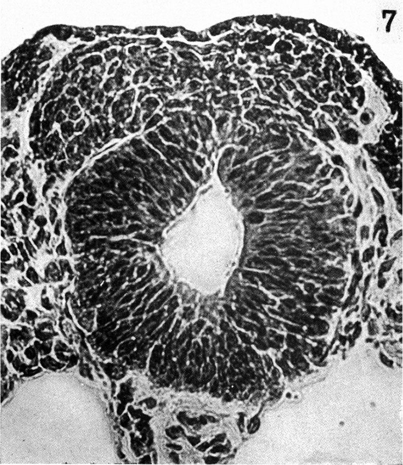 | 795 × 917 (242 KB) | Z8600021 (talk | contribs) | {{BaxterBoyd1939 figures}} |
You cannot overwrite this file.
File usage
The following 4 pages use this file: