File:BaxterBoyd1939-fig06.jpg: Difference between revisions
From Embryology
mNo edit summary |
mNo edit summary |
||
| Line 3: | Line 3: | ||
( x 500). Neural crest cells of the occipito-spinal group are to be seen in the angle between the neural tube and the somatic ectoderm. | ( x 500). Neural crest cells of the occipito-spinal group are to be seen in the angle between the neural tube and the somatic ectoderm. | ||
<br> | |||
{{Carnegie stage 10 links}} | |||
<br> | |||
{{Carnegie_stage_table_1}} | |||
<br> | |||
{{BaxterBoyd1939 figures}} | {{BaxterBoyd1939 figures}} | ||
[[Category:Neural Crest]][[Category:Somite]] | |||
[[Category:Neural Crest]] | |||
Latest revision as of 12:01, 28 May 2017
Fig. 6. Microphotograph of section at the level of the first somite in the Ten-Somite Human Embryo
( x 500). Neural crest cells of the occipito-spinal group are to be seen in the angle between the neural tube and the somatic ectoderm.
| Stage 10 Links: Week 4 | Gastrulation | Lecture | Practical | Image Gallery | Carnegie Embryos | Embryos | Category:Carnegie Stage 10 | Next Stage 11 |
| Historic Papers: 1910 | 1917 | 1926 | 1939 | 1943 | 1957 | 1985 |
| Week: | 1 | 2 | 3 | 4 | 5 | 6 | 7 | 8 |
| Carnegie stage: | 1 2 3 4 | 5 6 | 7 8 9 | 10 11 12 13 | 14 15 | 16 17 | 18 19 | 20 21 22 23 |
| Historic Disclaimer - information about historic embryology pages |
|---|
 |
- Links: Text-fig 1 | Text-fig 2 | Plate 1 | Fig 1 | Fig 2 | Fig 3 | Fig 4 | Plate 2 | Fig 5 | Fig 6 | Fig 7 | Baxter and Boyd 1939 | Historic Embryology Papers | Neural Crest Development | Carnegie stage 10
Reference
Baxter JS. and Boyd JD. Observations on the neural crest of a ten-somite human embryo. (1939) J Anat. 73: 318–326. PMID 17104759
Cite this page: Hill, M.A. (2024, April 18) Embryology BaxterBoyd1939-fig06.jpg. Retrieved from https://embryology.med.unsw.edu.au/embryology/index.php/File:BaxterBoyd1939-fig06.jpg
- © Dr Mark Hill 2024, UNSW Embryology ISBN: 978 0 7334 2609 4 - UNSW CRICOS Provider Code No. 00098G
File history
Click on a date/time to view the file as it appeared at that time.
| Date/Time | Thumbnail | Dimensions | User | Comment | |
|---|---|---|---|---|---|
| current | 22:11, 16 September 2015 | 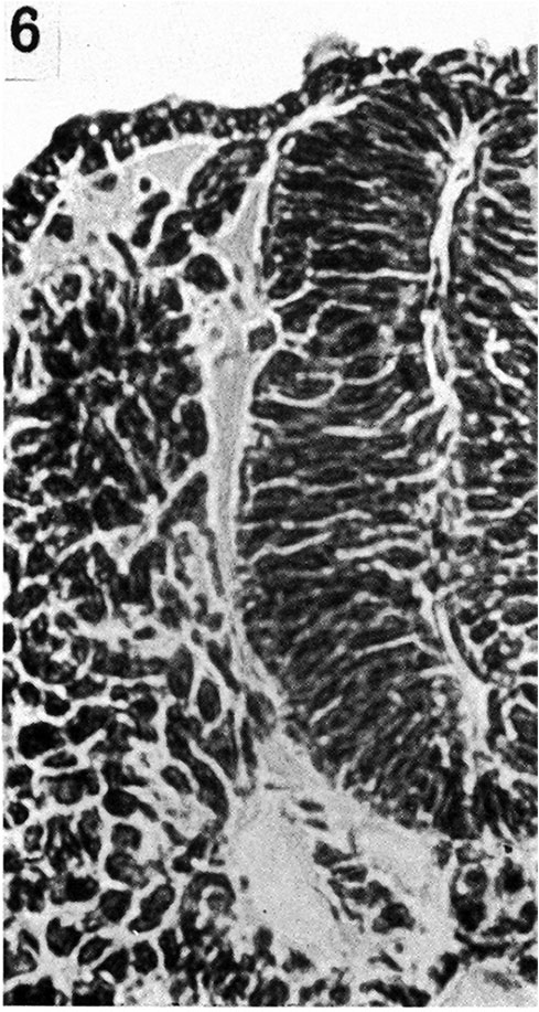 | 489 × 917 (141 KB) | Z8600021 (talk | contribs) | {{BaxterBoyd1939 figures}} |
You cannot overwrite this file.
File usage
The following 3 pages use this file: