File:Bartelmez1923 fig03.jpg: Difference between revisions
mNo edit summary |
mNo edit summary |
||
| (3 intermediate revisions by the same user not shown) | |||
| Line 3: | Line 3: | ||
Eternod’s nine somite embryo ‘DuGa’, based upon a complete series of tracings made by H. M. Evans. Magnified 200 diameters and reduced one half in reproduction. The neural tube is closed as far forward as the caudal end of the otic segment (rh.4). | Eternod’s nine somite embryo ‘DuGa’, based upon a complete series of tracings made by H. M. Evans. Magnified 200 diameters and reduced one half in reproduction. The neural tube is closed as far forward as the caudal end of the otic segment (rh.4). | ||
The broken lines at the level of the first somite represent the eight lost sections. The extent of the optic-neural crest primordia is indicated by stippling. ch.op.?, probably the chiasma ridge; c7'.neur., occipital neural crest; cr.n.rost., rostral division of the neural crest; dz'sc.ot., otic disc (outlined by dashes); mesen., midbrain; pr.a,c.fac., acoustico- facial primordium; p1'.op. , rostral end of the optic primordium which probably continues caudally to the region of crest proliferation; prosen., forebrain; rh.A1, the remains of the primary first hindbrain segment after the separation of the third definitive rhombomere (rh.8); rh.4 and 5, the otic and first postotic neuromeres; sac.br.I, the broken line indicates the extent of the first visceral pouch; sin.or., oral sinus; sul.cr., the clearly cut margin of the stipple indicates the extent of the ventricular cristal sulcus; s.1, probable first somite; thy., anlage of thyroid. The arrows indicate the levels of the sections of this embryo figured by me in 1922 (figs. 2c and 9b). | The broken lines at the level of the first somite represent the eight lost sections. The extent of the optic-neural crest primordia is indicated by stippling. ch.op.?, probably the chiasma ridge; c7'.neur., occipital neural crest; cr.n.rost., rostral division of the neural crest; dz'sc.ot., otic disc (outlined by dashes); mesen., midbrain; pr.a,c.fac., acoustico- facial primordium; p1'.op. , rostral end of the optic primordium which probably continues caudally to the region of crest proliferation; prosen., forebrain; rh.A1, the remains of the primary first hindbrain segment after the separation of the third definitive rhombomere (rh.8); rh.4 and 5, the otic and first postotic neuromeres; sac.br.I, the broken line indicates the extent of the first visceral pouch; sin.or., oral sinus; sul.cr., the clearly cut margin of the stipple indicates the extent of the ventricular cristal sulcus; s.1, probable first somite; thy., anlage of thyroid. The arrows indicate the levels of the sections of this embryo figured by me in 1922 (figs. 2c and 9b). Attention may be called here to a misprint in the legend to this figure. The section reproduced as figure 2c is number 21, not 41, of the series. | ||
{{Online Editor}} - Eternod’s ‘DuGa’ embryo, 9 somite embryo corresponds to [[Carnegie stage 10]] in [[Week 4]]. {{Ref-Eternod1899b}} | |||
<br> | |||
{{Carnegie stage 10 links}} | |||
<br> | |||
{{Carnegie_stage_table_1}} | |||
<br> | |||
{{Bartelmez1923 figures}} | {{Bartelmez1923 figures}} | ||
[[Category:Carnegie Stage 10]][[Category:Week 4]] | [[Category:Carnegie Stage 10]][[Category:Week 4]] | ||
Latest revision as of 11:57, 28 May 2017
Fig. 3 A projection reconstruction of the Head end of Eternod’s Nine Somite Embryo
Eternod’s nine somite embryo ‘DuGa’, based upon a complete series of tracings made by H. M. Evans. Magnified 200 diameters and reduced one half in reproduction. The neural tube is closed as far forward as the caudal end of the otic segment (rh.4).
The broken lines at the level of the first somite represent the eight lost sections. The extent of the optic-neural crest primordia is indicated by stippling. ch.op.?, probably the chiasma ridge; c7'.neur., occipital neural crest; cr.n.rost., rostral division of the neural crest; dz'sc.ot., otic disc (outlined by dashes); mesen., midbrain; pr.a,c.fac., acoustico- facial primordium; p1'.op. , rostral end of the optic primordium which probably continues caudally to the region of crest proliferation; prosen., forebrain; rh.A1, the remains of the primary first hindbrain segment after the separation of the third definitive rhombomere (rh.8); rh.4 and 5, the otic and first postotic neuromeres; sac.br.I, the broken line indicates the extent of the first visceral pouch; sin.or., oral sinus; sul.cr., the clearly cut margin of the stipple indicates the extent of the ventricular cristal sulcus; s.1, probable first somite; thy., anlage of thyroid. The arrows indicate the levels of the sections of this embryo figured by me in 1922 (figs. 2c and 9b). Attention may be called here to a misprint in the legend to this figure. The section reproduced as figure 2c is number 21, not 41, of the series.
Online Editor - Eternod’s ‘DuGa’ embryo, 9 somite embryo corresponds to Carnegie stage 10 in Week 4. Eternod ACF. II y a un canal notochordal dans l'embryon humain. (There is a notochordal channel in the human embryo) (1899) Anat. Anz., 16: 131-143.
| Stage 10 Links: Week 4 | Gastrulation | Lecture | Practical | Image Gallery | Carnegie Embryos | Embryos | Category:Carnegie Stage 10 | Next Stage 11 |
| Historic Papers: 1910 | 1917 | 1926 | 1939 | 1943 | 1957 | 1985 |
| Week: | 1 | 2 | 3 | 4 | 5 | 6 | 7 | 8 |
| Carnegie stage: | 1 2 3 4 | 5 6 | 7 8 9 | 10 11 12 13 | 14 15 | 16 17 | 18 19 | 20 21 22 23 |
| Historic Disclaimer - information about historic embryology pages |
|---|
 |
Reference
Bartelmez GW. The subdivisions of the neural folds in man. (1923) J. Comp. Neural., 35: 231-247.
Cite this page: Hill, M.A. (2024, April 16) Embryology Bartelmez1923 fig03.jpg. Retrieved from https://embryology.med.unsw.edu.au/embryology/index.php/File:Bartelmez1923_fig03.jpg
- © Dr Mark Hill 2024, UNSW Embryology ISBN: 978 0 7334 2609 4 - UNSW CRICOS Provider Code No. 00098G
File history
Click on a date/time to view the file as it appeared at that time.
| Date/Time | Thumbnail | Dimensions | User | Comment | |
|---|---|---|---|---|---|
| current | 22:46, 7 June 2016 | 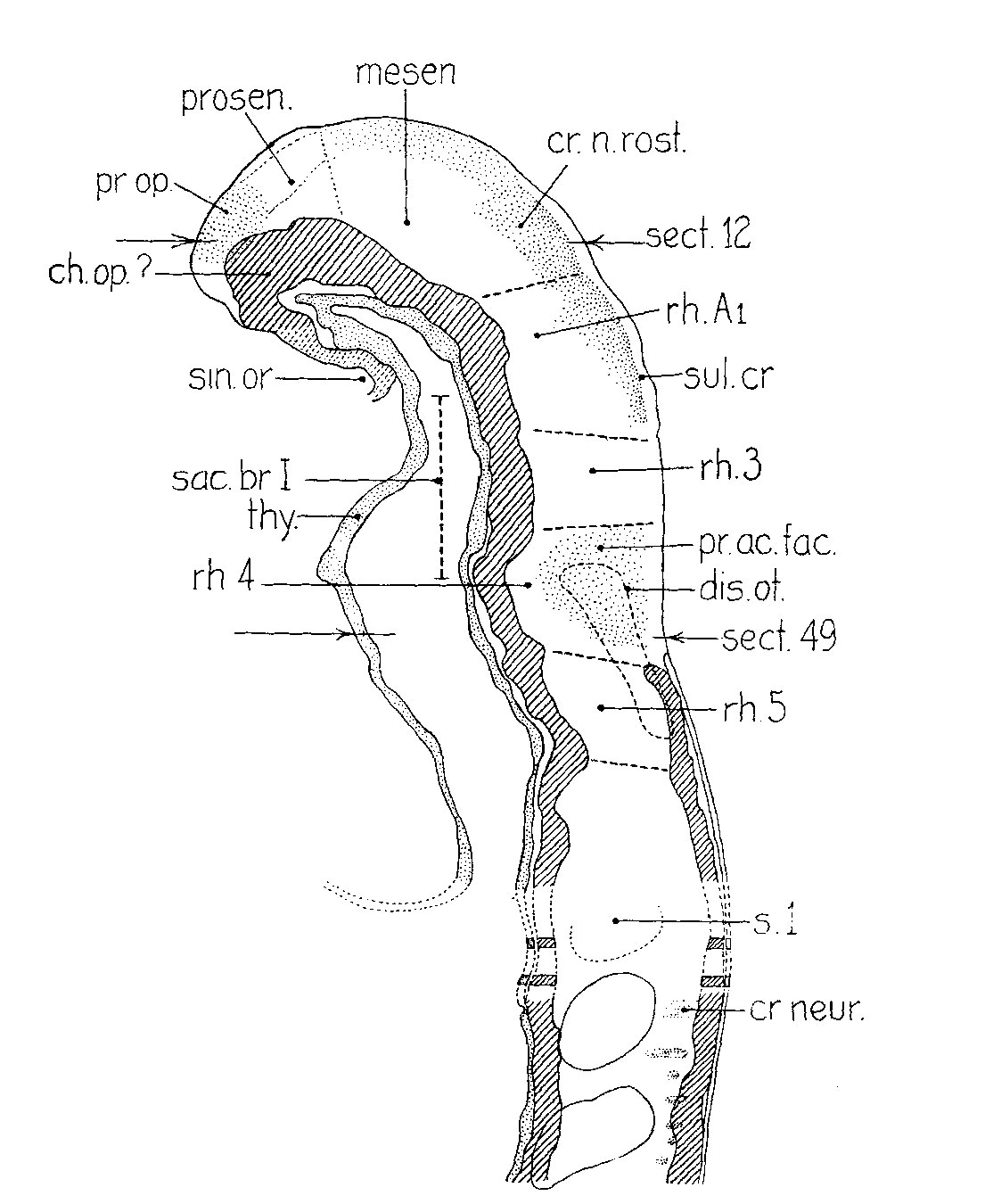 | 1,105 × 1,350 (202 KB) | Z8600021 (talk | contribs) |
You cannot overwrite this file.
File usage
The following 2 pages use this file: