File:BakerHookSeveringhaus1944 plate04.jpg

Original file (1,563 × 2,278 pixels, file size: 347 KB, MIME type: image/jpeg)
Plate 4
22 Cell column connecting a. villus at left to wall of Fallopian tube out of field at right. Three mitoses are present near cytotrophoblast of villus. Cells farthet removed from villus are larger and enclose empty cytoplasmic spaces from which glycogen has been removed. EH 265. 10 μm Hernatexylin and eosin. X 230.
23 Golgi apparatus and lohated nucleus of a small decidual cell at lower left. Case 14. X 1360.
24 Golgi apparatus in four large decidual cells. Case 14. X 960.
25 A cell island near two villi, the tropliohlast of which is seen at lower right and at the left. The island is not covered with syncytiuni and shows an early stage of degeneration at the center with loss of cell structure and a.ppeara:nce of small masses of librinoid. At left of island is a syncytial bud containing large vacuoles. The small black dots in the syncytium and loud are lipid droplets Case 13. x 240.
26 Connective tissue material enfoldecl by a large decidual cell and centinuous with the peri-cellular condensation. Benin. Severinghaus Altn1annMassen. 532.. Case 5. ,)< 1160.
27 Large deeidual cell with two mitochondria—laden processes partially surrounding dense connective tissue matrix. Mitochondria also accuniulated in the Golgi region above the nucleus. Case 2. x 1160.
28 Three large decidual cells and three small decidual cells, the latter with intensely chromatic nuclei and empty cytoplasmic space. The one at the right shows a. clump of blue granules. “Benin. Severinghaus Altmann-lrlasson. fin. Case 17. X 960.
29 Glycogen deposits in three small decidual cells. Large decitlual cell at upper right with some glycogen at its upper border. Picric-alcohol-formel. Best’s earmine. 5;; Case 5. X 960.
30 Pericellular eendensatiens around large decidual cells of zone cornpacta. Benin. Bielschowsky and Harris. hematexylin. Case 12. x 176.
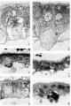
plate 1
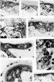
plate 2
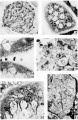
plate 3
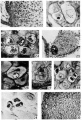
plate 4


Reference
Baker BL. Hook SJ. and Severinghaus AE. The cytological structure of the human chorionic villus and decidua parietalis. (1944) Amer. J Anat. 73(3): 291-325.
Cite this page: Hill, M.A. (2024, April 18) Embryology BakerHookSeveringhaus1944 plate04.jpg. Retrieved from https://embryology.med.unsw.edu.au/embryology/index.php/File:BakerHookSeveringhaus1944_plate04.jpg
- © Dr Mark Hill 2024, UNSW Embryology ISBN: 978 0 7334 2609 4 - UNSW CRICOS Provider Code No. 00098G
File history
Click on a date/time to view the file as it appeared at that time.
| Date/Time | Thumbnail | Dimensions | User | Comment | |
|---|---|---|---|---|---|
| current | 09:20, 2 February 2018 | | 1,563 × 2,278 (347 KB) | Z8600021 (talk | contribs) | |
| 09:20, 2 February 2018 | 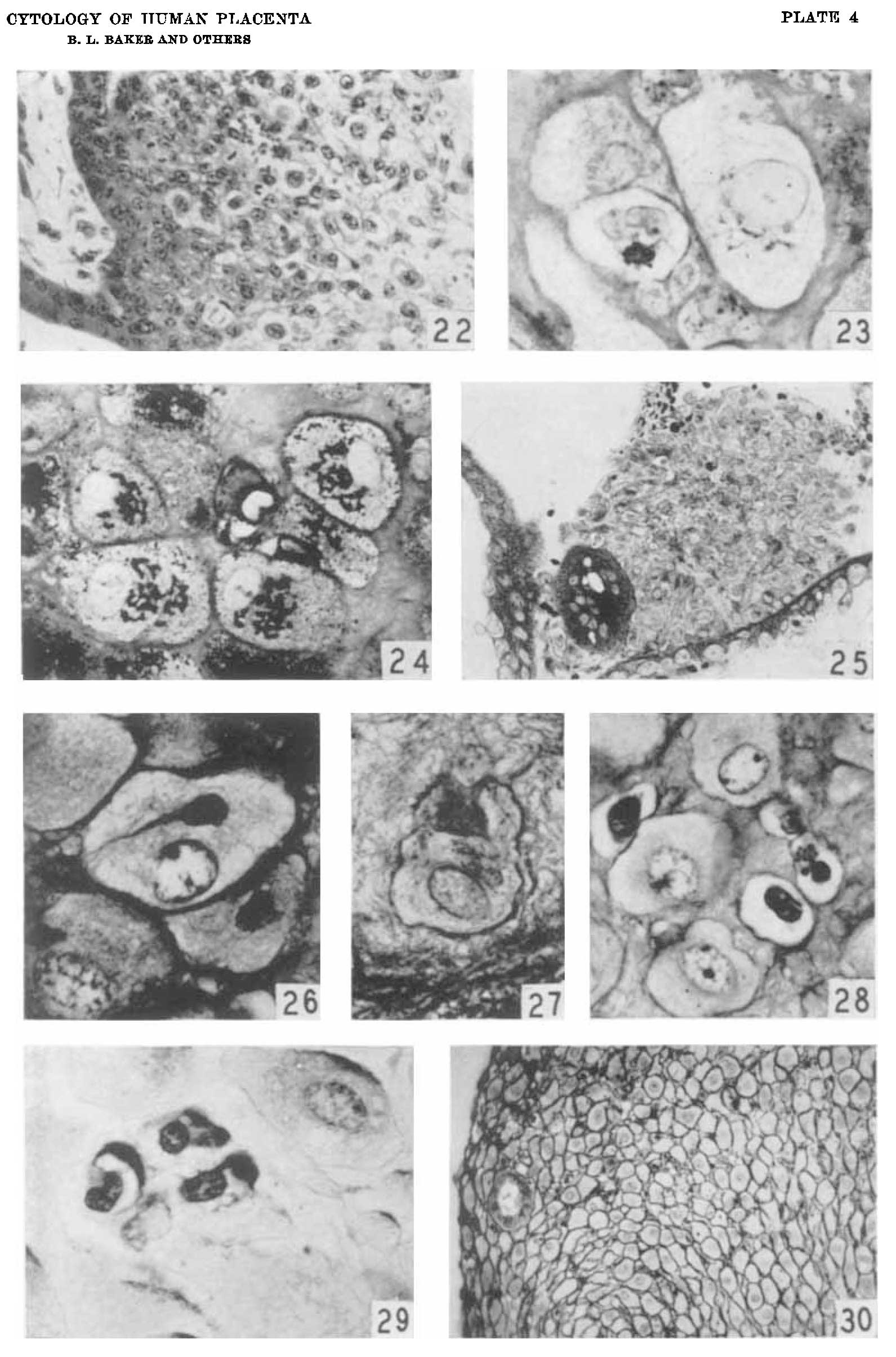 | 1,591 × 2,408 (301 KB) | Z8600021 (talk | contribs) | ==Plate 4== 22 Cell column connecting a. villus at left to wall of Fallopian tube out of field at right. Three mitoses are present near cytotrophoblast of villus. Cells farthet removed from villus are larger and enclose empty cytoplasmic spaces from w... |
You cannot overwrite this file.