File:BakerHookSeveringhaus1944 plate02.jpg

Original file (1,502 × 2,261 pixels, file size: 265 KB, MIME type: image/jpeg)
Plate 2
Unless otherwise stated, the figures are from 3 μm sections of Champy-fixed material stained by the Severinghaus Altmann—Masson technique; Golgi preparations were osmicated by the Sevoringliefus (Nassonov-Kolatschev) method before staining.
7 Loose Golgi apparatus of a large cytotrophoblast cell. Case 5. x 1160.
8 Compact Golgi apparatus containing vesicles in a small cytotrophoblast cell. At left of nucleus are brown lipid bodies. Not stained. (Jase 14. X 1740. 9 Golgi apparatus of a full-term cytotrophoblast. cell. Case 22. X 1160.
10 Full-term cytotrophoblast cell (arrow) with condensed cytoplasm in the Golgi region containing a few mitocliondria. Thin syncytium. covers this cell and on right side of villus above figure number is seen the low, scrubby brush border. The basement membrane is markedly thickened. Case 22. X 1.160.
11 0n the right (arrow) a cytotrophoblast cell in process of transformation to Sl}'I1(3__Ytl1.ll'l1 in which the cytoplasm is indistinguishable from that of the s_vnc.y'tium, but the ce.ll membrane is clearly visible above the nucleus. Mitochondria are clumped and briglitly stained. The syncytium shows some granules and superficial vacuoles with surrounding accumulation of niitochondria. Case 12. X 1400.
12 A syncytial bud with many mitochondria and a few small granules at the right of the most distal nucleus. Case 12. X 1160.
13 An area. from the edge of a cell island showing homogeneous fibrinoid at the top and a large granule-packed cell (arrow) with hypertrophied Golgi body near the bottom. Separation of the cells is indicated by the maternal erythrocytes scattered among them. Case 13. X 1160.
14 Seemingly viscous material on the surface of the villus holding maternal erythrocytes. Many of the latter stained partly red and partly yellow, or various shades of blue. A highly active syncytiiiin containing, in the supranuclcar zone, many lipid droplets (black) and granules (gray), the latter appearilig to be in various stages of liquefaction. Case 12. X 1160.
15 Thin, inactive syncytium of 5 months, containing only one or two minute granules at the left. Case 3. X 1160.
Reference
Baker BL. Hook SJ. and Severinghaus AE. The cytological structure of the human chorionic villus and decidua parietalis. (1944) Amer. J Anat. 73(3): 291-325.
Cite this page: Hill, M.A. (2024, April 20) Embryology BakerHookSeveringhaus1944 plate02.jpg. Retrieved from https://embryology.med.unsw.edu.au/embryology/index.php/File:BakerHookSeveringhaus1944_plate02.jpg
- © Dr Mark Hill 2024, UNSW Embryology ISBN: 978 0 7334 2609 4 - UNSW CRICOS Provider Code No. 00098G
File history
Click on a date/time to view the file as it appeared at that time.
| Date/Time | Thumbnail | Dimensions | User | Comment | |
|---|---|---|---|---|---|
| current | 09:42, 2 February 2018 | | 1,502 × 2,261 (265 KB) | Z8600021 (talk | contribs) | |
| 09:29, 2 February 2018 | 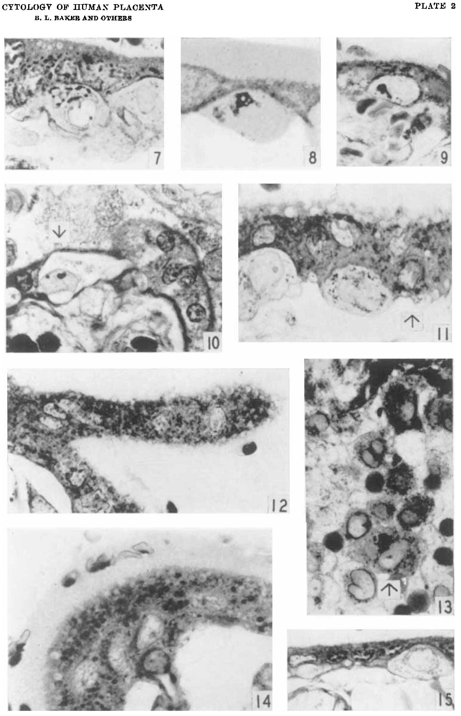 | 1,540 × 2,390 (216 KB) | Z8600021 (talk | contribs) |
You cannot overwrite this file.