File:BakerHookSeveringhaus1944 plate02.jpg: Difference between revisions
From Embryology
mNo edit summary |
mNo edit summary |
||
| Line 1: | Line 1: | ||
Unless otherwise stated, the figures are from 3 μm sections of Champy-fixed material stained by the Severinghaus Altmann—Masson technique; Golgi preparations were osmicated by the Sevoringliefus (Nassonov-Kolatschev) method before staining. | |||
===Reference=== | ===Reference=== | ||
{{Ref-BakerHookSeveringhaus1944}} | {{Ref-BakerHookSeveringhaus1944}} | ||
Revision as of 09:41, 2 February 2018
Unless otherwise stated, the figures are from 3 μm sections of Champy-fixed material stained by the Severinghaus Altmann—Masson technique; Golgi preparations were osmicated by the Sevoringliefus (Nassonov-Kolatschev) method before staining.
Reference
Baker BL. Hook SJ. and Severinghaus AE. The cytological structure of the human chorionic villus and decidua parietalis. (1944) Amer. J Anat. 73(3): 291-325.
Cite this page: Hill, M.A. (2024, April 25) Embryology BakerHookSeveringhaus1944 plate02.jpg. Retrieved from https://embryology.med.unsw.edu.au/embryology/index.php/File:BakerHookSeveringhaus1944_plate02.jpg
- © Dr Mark Hill 2024, UNSW Embryology ISBN: 978 0 7334 2609 4 - UNSW CRICOS Provider Code No. 00098G
File history
Click on a date/time to view the file as it appeared at that time.
| Date/Time | Thumbnail | Dimensions | User | Comment | |
|---|---|---|---|---|---|
| current | 09:42, 2 February 2018 |  | 1,502 × 2,261 (265 KB) | Z8600021 (talk | contribs) | |
| 09:29, 2 February 2018 | 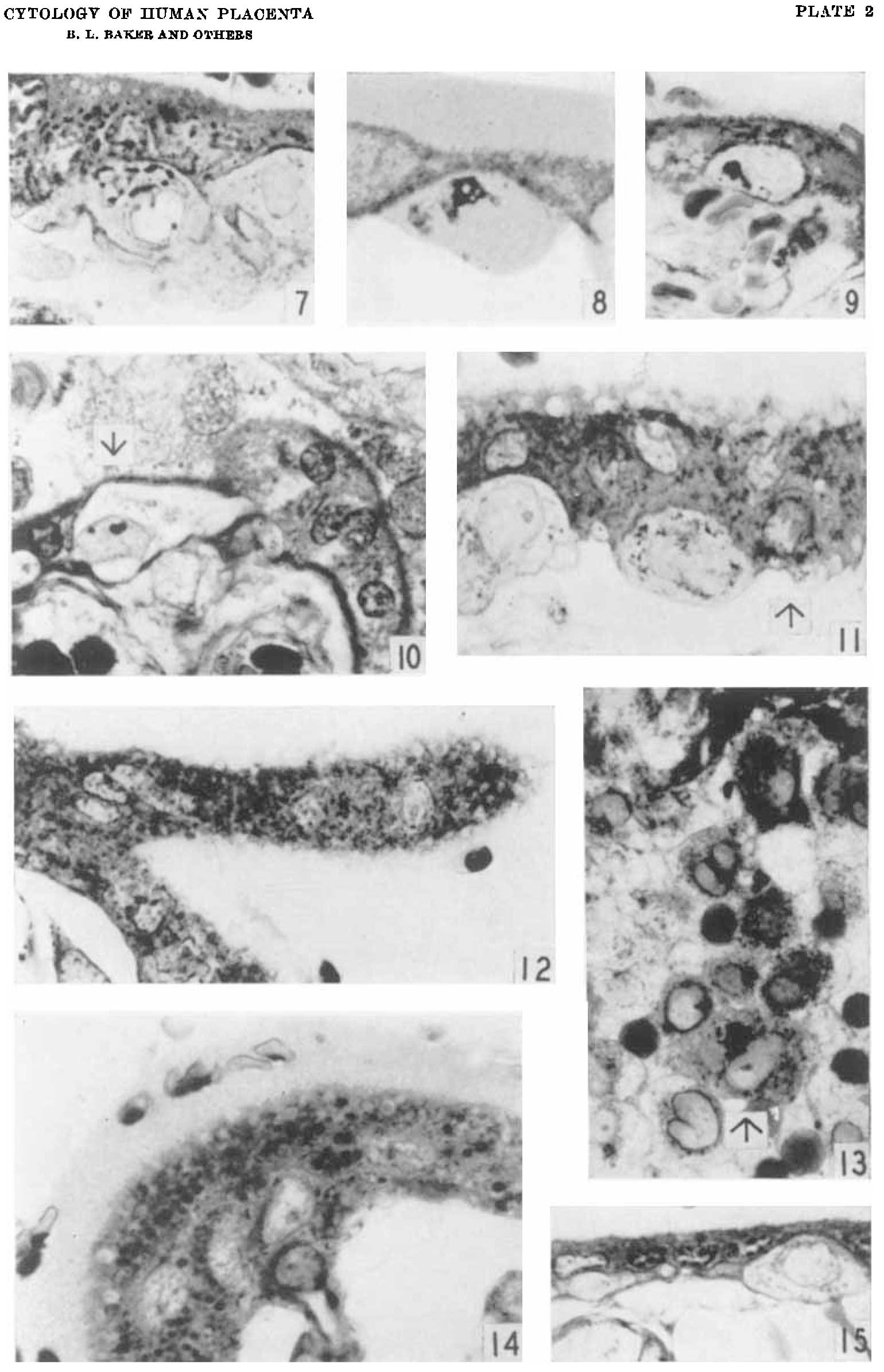 | 1,540 × 2,390 (216 KB) | Z8600021 (talk | contribs) |
You cannot overwrite this file.