File:Bailey502.jpg: Difference between revisions
({{Template:Bailey 1921 Figures}} Category:Coelom Category:Placenta) |
|||
| (2 intermediate revisions by one other user not shown) | |||
| Line 1: | Line 1: | ||
==Fig. 502. Placenta at birth, seen from the uterine side== | |||
Bonnet. | |||
Some of the chorionic villi float freely in the blood spaces of the maternal placenta floating villi; others are attached to the maternal tissue fastening villi. The villi are separated into larger and smaller groups or lobules by the growth of connective tissue septa from the maternal placenta down into the decidua basalis. These are known as placental septa, while the groups of chorionic villi are known as cotyledons (Figs. 500 and 502). | |||
Both decidual cells and chorionic villi are important from a diagnostic standpoint, as the finding of them in curettings or in a uterine discharge may be accepted as proof of pregnancy. | |||
During the early months of pregnancy first four months the decidua basalis has essentially the same structure as the decidua parietalis. Its surface epithelium disappears very early, perhaps even before the attachment of the ovum. The glandular elements and the connective tissue undergo the same changes as in the decidua parietalis and here also result in the differentiation of a compact layer and a spongy layer. Both layers are much thinner than in the decidua parietals. | |||
As already noted, connective tissue septa pass from the superficial layer of the decidua basalis down into the foetal placenta subdividing the latter into cotyledons. At the margin of the placenta the decidua basalis passes over into the thicker decidua parietalis and here the chorion is firmly attached to the decidua basalis. | |||
{{Template:Bailey 1921 Figures}} | {{Template:Bailey 1921 Figures}} | ||
[[Category: | [[Category:Human]] [[Category:Placenta]] [[Category:Birth]] [[Category:Third Trimester]] | ||
Latest revision as of 11:44, 6 June 2012
Fig. 502. Placenta at birth, seen from the uterine side
Bonnet.
Some of the chorionic villi float freely in the blood spaces of the maternal placenta floating villi; others are attached to the maternal tissue fastening villi. The villi are separated into larger and smaller groups or lobules by the growth of connective tissue septa from the maternal placenta down into the decidua basalis. These are known as placental septa, while the groups of chorionic villi are known as cotyledons (Figs. 500 and 502).
Both decidual cells and chorionic villi are important from a diagnostic standpoint, as the finding of them in curettings or in a uterine discharge may be accepted as proof of pregnancy.
During the early months of pregnancy first four months the decidua basalis has essentially the same structure as the decidua parietalis. Its surface epithelium disappears very early, perhaps even before the attachment of the ovum. The glandular elements and the connective tissue undergo the same changes as in the decidua parietalis and here also result in the differentiation of a compact layer and a spongy layer. Both layers are much thinner than in the decidua parietals.
As already noted, connective tissue septa pass from the superficial layer of the decidua basalis down into the foetal placenta subdividing the latter into cotyledons. At the margin of the placenta the decidua basalis passes over into the thicker decidua parietalis and here the chorion is firmly attached to the decidua basalis.
- Text-Book of Embryology: Germ cells | Maturation | Fertilization | Amphioxus | Frog | Chick | Mammalian | External body form | Connective tissues and skeletal | Vascular | Muscular | Alimentary tube and organs | Respiratory | Coelom, Diaphragm and Mesenteries | Urogenital | Integumentary | Nervous System | Special Sense | Foetal Membranes | Teratogenesis | Gallery of All Figures
| Historic Disclaimer - information about historic embryology pages |
|---|
 |
Reference
Bailey FR. and Miller AM. Text-Book of Embryology (1921) New York: William Wood and Co.
Cite this page: Hill, M.A. (2024, April 16) Embryology Bailey502.jpg. Retrieved from https://embryology.med.unsw.edu.au/embryology/index.php/File:Bailey502.jpg
- © Dr Mark Hill 2024, UNSW Embryology ISBN: 978 0 7334 2609 4 - UNSW CRICOS Provider Code No. 00098G
File history
Click on a date/time to view the file as it appeared at that time.
| Date/Time | Thumbnail | Dimensions | User | Comment | |
|---|---|---|---|---|---|
| current | 23:47, 1 February 2011 | 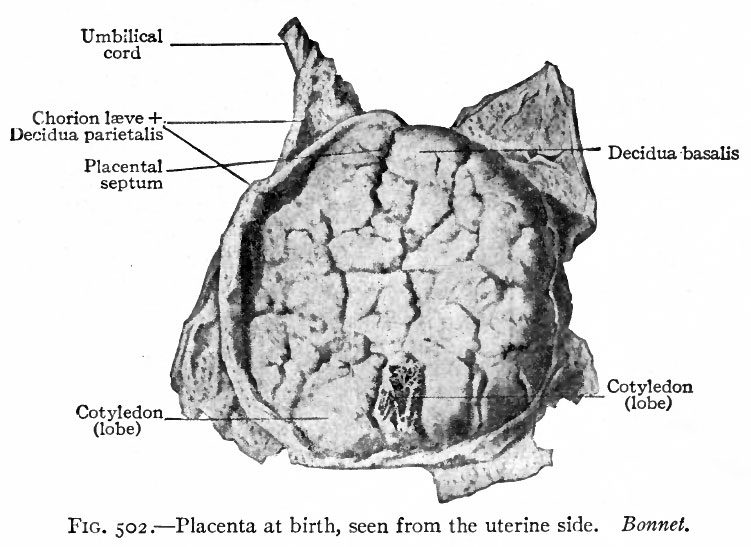 | 751 × 547 (74 KB) | S8600021 (talk | contribs) | {{Template:Bailey 1921 Figures}} Category:Coelom Category:Placenta |
You cannot overwrite this file.