File:Bailey463.jpg
Bailey463.jpg (679 × 345 pixels, file size: 52 KB, MIME type: image/jpeg)
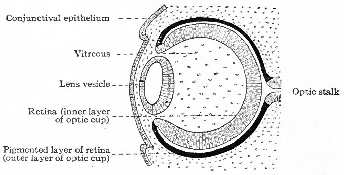
Fig. 463. Diagram of developing lens and optic cup
Duval.
The cells of the inner wall of the lens vesicle have begun to elongate to form lens fibers. The epithelium over the lens is the anlage of the corneal epithelium. The mesodermal tissue between the latter and the anterior wall of the lens vesicle is the anlage of the substantia propria cornea.
At first the lens vesicle is connected with the surface ectoderm, but about the eighth week a thin layer of mesoderm grows in between the lens vesicle and the surface ectoderm, completely separating them (Fig. 463). The ingrowth of the lens vesicle against the outgrowing optic vesicle has the effect as though a small hard ball (the lens vesicle) had been pressed into a larger soft ball (the optic vesicle) (Fig. 464) . The lens vesicle pushes the outer wall of the optic vesicle in against the inner wall, the optic vesicle thus becoming transformed into the two-layered optic cup (Figs. 462, 463).
- Text-Book of Embryology: Germ cells | Maturation | Fertilization | Amphioxus | Frog | Chick | Mammalian | External body form | Connective tissues and skeletal | Vascular | Muscular | Alimentary tube and organs | Respiratory | Coelom, Diaphragm and Mesenteries | Urogenital | Integumentary | Nervous System | Special Sense | Foetal Membranes | Teratogenesis | Gallery of All Figures
| Historic Disclaimer - information about historic embryology pages |
|---|
 |
Reference
Bailey FR. and Miller AM. Text-Book of Embryology (1921) New York: William Wood and Co.
Cite this page: Hill, M.A. (2024, April 24) Embryology Bailey463.jpg. Retrieved from https://embryology.med.unsw.edu.au/embryology/index.php/File:Bailey463.jpg
- © Dr Mark Hill 2024, UNSW Embryology ISBN: 978 0 7334 2609 4 - UNSW CRICOS Provider Code No. 00098G
File history
Click on a date/time to view the file as it appeared at that time.
| Date/Time | Thumbnail | Dimensions | User | Comment | |
|---|---|---|---|---|---|
| current | 13:46, 1 February 2011 | | 679 × 345 (52 KB) | S8600021 (talk | contribs) |
You cannot overwrite this file.