File:Bailey462.jpg
Bailey462.jpg (463 × 411 pixels, file size: 45 KB, MIME type: image/jpeg)
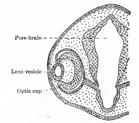
Fig. 462. Showing somewhat later stage in development of optic cup and lens than is shown in Fig. 461
Duval.
The thickened portion of ectoderm is known as the lens area (Fig. 460). The latter next becomes depressed against the outet surface of the optic vesicle forming a distinct lens invagination (Fig. 461). This becomes cup-shaped and then its edges come together and fuse, thus forming the lens vesicle (Fig. 462). At first the lens vesicle is connected with the surface ectoderm, but about the eighth week a thin layer of mesoderm grows in between the lens vesicle and the surface ectoderm, completely separating them (Fig. 463).
- Text-Book of Embryology: Germ cells | Maturation | Fertilization | Amphioxus | Frog | Chick | Mammalian | External body form | Connective tissues and skeletal | Vascular | Muscular | Alimentary tube and organs | Respiratory | Coelom, Diaphragm and Mesenteries | Urogenital | Integumentary | Nervous System | Special Sense | Foetal Membranes | Teratogenesis | Gallery of All Figures
| Historic Disclaimer - information about historic embryology pages |
|---|
 |
Reference
Bailey FR. and Miller AM. Text-Book of Embryology (1921) New York: William Wood and Co.
Cite this page: Hill, M.A. (2024, April 25) Embryology Bailey462.jpg. Retrieved from https://embryology.med.unsw.edu.au/embryology/index.php/File:Bailey462.jpg
- © Dr Mark Hill 2024, UNSW Embryology ISBN: 978 0 7334 2609 4 - UNSW CRICOS Provider Code No. 00098G
File history
Click on a date/time to view the file as it appeared at that time.
| Date/Time | Thumbnail | Dimensions | User | Comment | |
|---|---|---|---|---|---|
| current | 13:46, 1 February 2011 | | 463 × 411 (45 KB) | S8600021 (talk | contribs) |
You cannot overwrite this file.
File usage
The following 4 pages use this file: