File:Bailey460.jpg
Bailey460.jpg (718 × 423 pixels, file size: 55 KB, MIME type: image/jpeg)
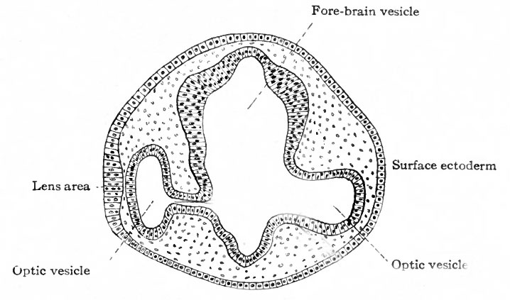
Fig. 460. Section through head of chick of two days' incubation
Duval.
The formation of the optic vesicle and stalk appears to be somewhat more advanced on the left than on the right.
The first indication of eye formation is found in the chick at the beginning of the second day of incubation ; in the human embryo, at what has been estimated as about the second or third week. At this stage the neural canal is not yet completely closed in and its anterior end shows three primary brain vesicles (p. 440, Fig. 497). The anlagen of the eyes first appear as bilaterally symmetrical evaginations from the lateral walls of the fore-brain vesicle (Figs. 459 and 460), and are at first large in proportion to the brain vesicle itself. When first formed, the optic evagination opens widely into the fore-brain vesicle (Fig. 460, right side), but as the distal part of the evagination expands more rapidly than the proximal part, there soon results a spheroidal optic vesicle attached to the fore-brain by the narrow optic stalk (Fig. 460, left side) . Through the latter the cavity of the optic vesicle and the cavity of the fore-brain are in communication. With the development of the hemispheres, that part of the brain to which the optic stalks are attached becomes the inter-brain (diencephalon).
- Text-Book of Embryology: Germ cells | Maturation | Fertilization | Amphioxus | Frog | Chick | Mammalian | External body form | Connective tissues and skeletal | Vascular | Muscular | Alimentary tube and organs | Respiratory | Coelom, Diaphragm and Mesenteries | Urogenital | Integumentary | Nervous System | Special Sense | Foetal Membranes | Teratogenesis | Gallery of All Figures
| Historic Disclaimer - information about historic embryology pages |
|---|
 |
Reference
Bailey FR. and Miller AM. Text-Book of Embryology (1921) New York: William Wood and Co.
Cite this page: Hill, M.A. (2024, April 24) Embryology Bailey460.jpg. Retrieved from https://embryology.med.unsw.edu.au/embryology/index.php/File:Bailey460.jpg
- © Dr Mark Hill 2024, UNSW Embryology ISBN: 978 0 7334 2609 4 - UNSW CRICOS Provider Code No. 00098G
File history
Click on a date/time to view the file as it appeared at that time.
| Date/Time | Thumbnail | Dimensions | User | Comment | |
|---|---|---|---|---|---|
| current | 01:59, 30 January 2011 | | 718 × 423 (55 KB) | S8600021 (talk | contribs) | {{Template:Bailey 1921 Figures}} Category:Neural |
You cannot overwrite this file.
File usage
The following 4 pages use this file: