File:Bailey458-459.jpg
Bailey458-459.jpg (741 × 386 pixels, file size: 42 KB, MIME type: image/jpeg)
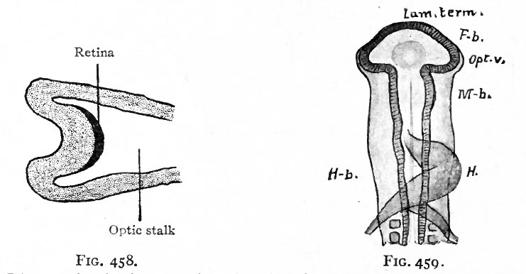
Fig. 458. Diagram showing, location of the (dark) optic area (see Fig. 457) after the beginning of the formation of the optic cup and optic stalk
Lange.
Fig. 459. Dorsal view of head of chick of 58 hours' incubation
Mihalkovics.
Lam. term, lamina terminalis; Fb., fore-brain; Opt. v., optic vesicle; M. b., mid-brain; H.b., hind or rhombic brain; H., heart.
In the eye the cell bodies of these neurones are located in the retina, but the area of ectoderm from which the retina develops first occupies a position along the neural crest analogous to that occupied by the anlagen of the spinal and cranial ganglia. In the case of the retina this area, instead of becoming split off in the closure of the neural canal, becomes folded into the canal and later pushed out toward the surface in the optic evagination (Figs. 450, 457, 458).
The first indication of eye formation is found in the chick at the beginning of the second day of incubation ; in the human embryo, at what has been estimated as about the second or third week. At this stage the neural canal is not yet completely closed in and its anterior end shows three primary brain vesicles (p. 440, Fig. 497). The anlagen of the eyes first appear as bilaterally symmetrical evaginations from the lateral walls of the fore-brain vesicle (Figs. 459 and 460), and are at first large in proportion to the brain vesicle itself. When first formed, the optic evagination opens widely into the fore-brain vesicle (Fig. 460, right side), but as the distal part of the evagination expands more rapidly than the proximal part, there soon results a spheroidal optic vesicle attached to the fore-brain by the narrow optic stalk (Fig. 460, left side) .
- Text-Book of Embryology: Germ cells | Maturation | Fertilization | Amphioxus | Frog | Chick | Mammalian | External body form | Connective tissues and skeletal | Vascular | Muscular | Alimentary tube and organs | Respiratory | Coelom, Diaphragm and Mesenteries | Urogenital | Integumentary | Nervous System | Special Sense | Foetal Membranes | Teratogenesis | Gallery of All Figures
| Historic Disclaimer - information about historic embryology pages |
|---|
 |
Reference
Bailey FR. and Miller AM. Text-Book of Embryology (1921) New York: William Wood and Co.
Cite this page: Hill, M.A. (2024, April 23) Embryology Bailey458-459.jpg. Retrieved from https://embryology.med.unsw.edu.au/embryology/index.php/File:Bailey458-459.jpg
- © Dr Mark Hill 2024, UNSW Embryology ISBN: 978 0 7334 2609 4 - UNSW CRICOS Provider Code No. 00098G
File history
Click on a date/time to view the file as it appeared at that time.
| Date/Time | Thumbnail | Dimensions | User | Comment | |
|---|---|---|---|---|---|
| current | 01:59, 30 January 2011 | | 741 × 386 (42 KB) | S8600021 (talk | contribs) |
You cannot overwrite this file.
File usage
The following 4 pages use this file: