File:Bailey456.jpg
Bailey456.jpg (591 × 168 pixels, file size: 20 KB, MIME type: image/jpeg)
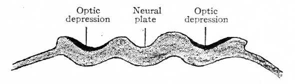
Fig. 456. Diagram showing location of optic areas before the closure of the neural groove
Modified from Lange.
The receptive mechanisms of all the general and special sense organs are derived from the ectoderm. With the single exception of the eye, all develop as direct specializations of the ectoderm in the form of the various neuro-epithelia. The eye is peculiar among the sense organs in that its receptive cells are not derived directly from surface ectoderm, but only indirectly from the ectoderm after it has become folded in to form the neural canal. The neuro-epithelium of the eye develops as a direct outgrowth from the central nervous system. The retina is a modified part of the brain; the optic nerves correspond to central nervous system fiber tracts. Of the accessory optic structures, the lens, the epithelium of the lids and conjunctiva, the eyelashes, the Meibomian glands and the epithelium of the lacrymal apparatus arc of ectodermic origin; the coats of the eye, the sclera and chorioid, and parts of their modified anterior extensions, the cornea, ciliary body and iris, are of mesodermic origin. In the sensory divisions of the other spinal and cranial nerves, with the exception of the olfactory, the cell bodies of the neurones which serve to connect the receptive mechanisms with the brain and cord are located in parts (the sensory ganglia of the cranial and spinal nerves) which have become separated from the crests of the neural folds as the latter fuse to form the neural canal. In the eye the cell bodies of these neurones are located in the retina, but the area of ectoderm from which the retina develops first occupies a position along the neural crest analogous to that occupied by the anlagen of the spinal and cranial ganglia. In the case of the retina this area, instead of becoming split off in the closure of the neural canal, becomes folded into the canal and later pushed out toward the surface in the optic evagination (Figs. 456, 457, 458).
- Text-Book of Embryology: Germ cells | Maturation | Fertilization | Amphioxus | Frog | Chick | Mammalian | External body form | Connective tissues and skeletal | Vascular | Muscular | Alimentary tube and organs | Respiratory | Coelom, Diaphragm and Mesenteries | Urogenital | Integumentary | Nervous System | Special Sense | Foetal Membranes | Teratogenesis | Gallery of All Figures
| Historic Disclaimer - information about historic embryology pages |
|---|
 |
Reference
Bailey FR. and Miller AM. Text-Book of Embryology (1921) New York: William Wood and Co.
Cite this page: Hill, M.A. (2024, April 20) Embryology Bailey456.jpg. Retrieved from https://embryology.med.unsw.edu.au/embryology/index.php/File:Bailey456.jpg
- © Dr Mark Hill 2024, UNSW Embryology ISBN: 978 0 7334 2609 4 - UNSW CRICOS Provider Code No. 00098G
File history
Click on a date/time to view the file as it appeared at that time.
| Date/Time | Thumbnail | Dimensions | User | Comment | |
|---|---|---|---|---|---|
| current | 01:58, 30 January 2011 | 591 × 168 (20 KB) | S8600021 (talk | contribs) | ||
| 01:57, 30 January 2011 |  | 593 × 509 (57 KB) | S8600021 (talk | contribs) | ||
| 01:57, 30 January 2011 |  | 809 × 388 (45 KB) | S8600021 (talk | contribs) | {{Template:Bailey 1921 Figures}} Category:Neural |
You cannot overwrite this file.
File usage
The following 4 pages use this file: