File:Bailey421.jpg: Difference between revisions
({{Template:Bailey 1921 Figures}} Category:Neural) |
mNo edit summary |
||
| (One intermediate revision by one other user not shown) | |||
| Line 1: | Line 1: | ||
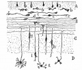
==Fig. 421. Scheme showing the various stages of position and form in the differentiation of granule cells from the outer granular layer== | |||
[[Category:Neural]] | [[Embryology History - Santiago Ramón y Cajal|Cajal]] drawing. | ||
A, Layer of undifferentiated cells; B, layer of cells in horizontal bipolar stage; C, partly formed molecular (plexiform) layer; D, granular layer; b, beginning differentiation of granule cells; c, cells in mo no polar stage; d, cells in bipolar stage; e,f, beginning of descending dendrite and of unipolarization of cell; g,h, i, different stages of unipolarization or formation of single process connecting with the original two processes; j, cell showing differentiating and completed dendrites; k, fully formed granule cell. | |||
{{Bailey 1921 Figures}} | |||
[[Category:Neural]] [[Category:Cajal]][[Category:Cerebellum]] | |||
Latest revision as of 08:44, 23 June 2015
Fig. 421. Scheme showing the various stages of position and form in the differentiation of granule cells from the outer granular layer
Cajal drawing.
A, Layer of undifferentiated cells; B, layer of cells in horizontal bipolar stage; C, partly formed molecular (plexiform) layer; D, granular layer; b, beginning differentiation of granule cells; c, cells in mo no polar stage; d, cells in bipolar stage; e,f, beginning of descending dendrite and of unipolarization of cell; g,h, i, different stages of unipolarization or formation of single process connecting with the original two processes; j, cell showing differentiating and completed dendrites; k, fully formed granule cell.
- Text-Book of Embryology: Germ cells | Maturation | Fertilization | Amphioxus | Frog | Chick | Mammalian | External body form | Connective tissues and skeletal | Vascular | Muscular | Alimentary tube and organs | Respiratory | Coelom, Diaphragm and Mesenteries | Urogenital | Integumentary | Nervous System | Special Sense | Foetal Membranes | Teratogenesis | Gallery of All Figures
| Historic Disclaimer - information about historic embryology pages |
|---|
| Pages where the terms "Historic" (textbooks, papers, people, recommendations) appear on this site, and sections within pages where this disclaimer appears, indicate that the content and scientific understanding are specific to the time of publication. This means that while some scientific descriptions are still accurate, the terminology and interpretation of the developmental mechanisms reflect the understanding at the time of original publication and those of the preceding periods, these terms, interpretations and recommendations may not reflect our current scientific understanding. (More? Embryology History | Historic Embryology Papers) |
Reference
Bailey FR. and Miller AM. Text-Book of Embryology (1921) New York: William Wood and Co.
Cite this page: Hill, M.A. (2024, April 19) Embryology Bailey421.jpg. Retrieved from https://embryology.med.unsw.edu.au/embryology/index.php/File:Bailey421.jpg
- © Dr Mark Hill 2024, UNSW Embryology ISBN: 978 0 7334 2609 4 - UNSW CRICOS Provider Code No. 00098G
File history
Click on a date/time to view the file as it appeared at that time.
| Date/Time | Thumbnail | Dimensions | User | Comment | |
|---|---|---|---|---|---|
| current | 01:34, 30 January 2011 |  | 625 × 538 (81 KB) | S8600021 (talk | contribs) | {{Template:Bailey 1921 Figures}} Category:Neural |
You cannot overwrite this file.
