File:Bailey387.jpg: Difference between revisions
From Embryology
No edit summary |
mNo edit summary |
||
| (One intermediate revision by the same user not shown) | |||
| Line 1: | Line 1: | ||
==Fig. 387. Dorsal portion of the lumbar cord of a chick embryo of three days== | ==Fig. 387. Dorsal portion of the lumbar cord of a chick embryo of three days== | ||
[[Embryology History - Santiago Ramón y Cajal|Cajal]] drawing. | |||
A, B, Cells in the apolar stage with fibrillogenous zones; B shows transition to the bipolar stage; E, further advanced bipolar cell; G, cells in monopolar stage or neuroblasts of His; a, giant cone of growth. These cells have migrated to the outer part of the nuclear layer, thereby forming the beginning of the mantle layer. | A, B, Cells in the apolar stage with fibrillogenous zones; B shows transition to the bipolar stage; E, further advanced bipolar cell; G, cells in monopolar stage or neuroblasts of His; a, giant cone of growth. These cells have migrated to the outer part of the nuclear layer, thereby forming the beginning of the mantle layer. | ||
{{ | {{Bailey 1921 Figures}} | ||
[[Category:Chicken]] [[Category:Neural]] [[Category:Cajal]] | [[Category:Chicken]] [[Category:Neural]] [[Category:Cajal]] | ||
Latest revision as of 08:39, 23 June 2015
Fig. 387. Dorsal portion of the lumbar cord of a chick embryo of three days
Cajal drawing.
A, B, Cells in the apolar stage with fibrillogenous zones; B shows transition to the bipolar stage; E, further advanced bipolar cell; G, cells in monopolar stage or neuroblasts of His; a, giant cone of growth. These cells have migrated to the outer part of the nuclear layer, thereby forming the beginning of the mantle layer.
- Text-Book of Embryology: Germ cells | Maturation | Fertilization | Amphioxus | Frog | Chick | Mammalian | External body form | Connective tissues and skeletal | Vascular | Muscular | Alimentary tube and organs | Respiratory | Coelom, Diaphragm and Mesenteries | Urogenital | Integumentary | Nervous System | Special Sense | Foetal Membranes | Teratogenesis | Gallery of All Figures
| Historic Disclaimer - information about historic embryology pages |
|---|
 |
Reference
Bailey FR. and Miller AM. Text-Book of Embryology (1921) New York: William Wood and Co.
Cite this page: Hill, M.A. (2024, April 18) Embryology Bailey387.jpg. Retrieved from https://embryology.med.unsw.edu.au/embryology/index.php/File:Bailey387.jpg
- © Dr Mark Hill 2024, UNSW Embryology ISBN: 978 0 7334 2609 4 - UNSW CRICOS Provider Code No. 00098G
File history
Click on a date/time to view the file as it appeared at that time.
| Date/Time | Thumbnail | Dimensions | User | Comment | |
|---|---|---|---|---|---|
| current | 00:36, 30 January 2011 | 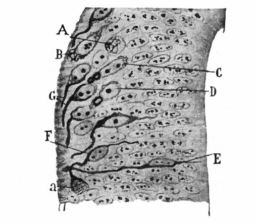 | 507 × 442 (50 KB) | S8600021 (talk | contribs) | ==Fig. 387. Dorsal portion of the lumbar cord of a chick embryo of three days== Cafal. A, B, Cells in the apolar stage with fibrillogenous zones; B shows transition to the bipolar stage; E, further advanced bipolar cell; G, cells in monopolar stage or |
You cannot overwrite this file.
File usage
The following 4 pages use this file: