File:Bailey322.jpg: Difference between revisions
From Embryology
({{Template:Bailey 1921 Figures}} Category:Human Category:Renal) |
m (→Fig. 322. From a model of the cloaca and the surrounding structures in a human embryo of 6.5 mm) |
||
| (3 intermediate revisions by 2 users not shown) | |||
| Line 1: | Line 1: | ||
{{ | ==Fig. 322. From a model of the cloaca and the surrounding structures in a human embryo of 6.5 mm== | ||
{{Keibel}} | |||
* The portion of the gut immediately caudal to the attachment of the allantoic duct becomes dilated to form the cloaca which at first is a blind sac, its cavity being separated from the outer surface of the embryo by the cloacal membrane (Fig. 322). | |||
* The openings of the mesonephric ducts, which primarily were situated in the lateral cloacal wall (p. 359), are situated after the separation in the dorso-lateral wall of the urogenital sinus (compare Figs. [[:File:Bailey322.jpg|322]], [[:File:Bailey323.jpg|323]], [[:File:Bailey324.jpg|324]]). | |||
:Link: [[Book_-_Text-Book_of_Embryology_15#Fig322|Figure in Text]] | |||
{{Bailey 1921 Figures}} | |||
[[Category:Human]] [[Category:Renal]] | [[Category:Human]] [[Category:Renal]] | ||
Latest revision as of 13:50, 31 May 2016
Fig. 322. From a model of the cloaca and the surrounding structures in a human embryo of 6.5 mm
Franz Keibel (1861-1929)
- The portion of the gut immediately caudal to the attachment of the allantoic duct becomes dilated to form the cloaca which at first is a blind sac, its cavity being separated from the outer surface of the embryo by the cloacal membrane (Fig. 322).
- The openings of the mesonephric ducts, which primarily were situated in the lateral cloacal wall (p. 359), are situated after the separation in the dorso-lateral wall of the urogenital sinus (compare Figs. 322, 323, 324).
- Link: Figure in Text
- Text-Book of Embryology: Germ cells | Maturation | Fertilization | Amphioxus | Frog | Chick | Mammalian | External body form | Connective tissues and skeletal | Vascular | Muscular | Alimentary tube and organs | Respiratory | Coelom, Diaphragm and Mesenteries | Urogenital | Integumentary | Nervous System | Special Sense | Foetal Membranes | Teratogenesis | Gallery of All Figures
| Historic Disclaimer - information about historic embryology pages |
|---|
 |
Reference
Bailey FR. and Miller AM. Text-Book of Embryology (1921) New York: William Wood and Co.
Cite this page: Hill, M.A. (2024, April 24) Embryology Bailey322.jpg. Retrieved from https://embryology.med.unsw.edu.au/embryology/index.php/File:Bailey322.jpg
- © Dr Mark Hill 2024, UNSW Embryology ISBN: 978 0 7334 2609 4 - UNSW CRICOS Provider Code No. 00098G
File history
Click on a date/time to view the file as it appeared at that time.
| Date/Time | Thumbnail | Dimensions | User | Comment | |
|---|---|---|---|---|---|
| current | 12:00, 25 January 2011 | 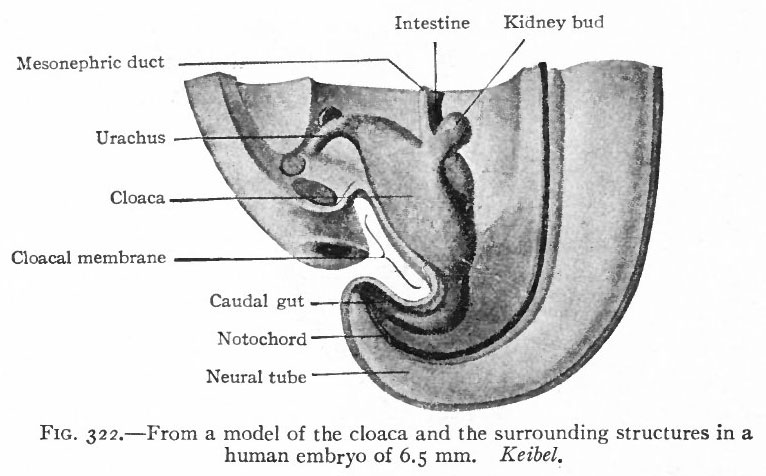 | 766 × 476 (62 KB) | S8600021 (talk | contribs) | {{Template:Bailey 1921 Figures}} Category:Human Category:Renal |
You cannot overwrite this file.
File usage
The following 3 pages use this file: