File:Bailey301-303.jpg

Original file (916 × 1,097 pixels, file size: 172 KB, MIME type: image/jpeg)
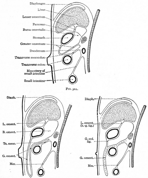
Figs. 301, 302 and 303. Diagrams showing stages in the development of the bursa omentalis, the greater omentum, and the fusion of the latter with the transverse mesocolon
Diagrams represent sagittal sections (For explanation of lettering in Figs. 302 and 303 see Fig. 301).
From the third month on, the greater omentum becomes larger and gradually extends toward the ventral abdominal wall, over the transverse colon, and then caudally between the body wall and the small intestine (Figs. 301 and 302). The portion between the body wall and intestine encloses merely a flat cavity continuous with the larger cavity dorsal to the stomach.
From the fourth month on, the omentum fuses with certain other structures and becomes less free. The dorsal lamella fuses with the dorsal body wall on the left side and with the transverse mesocolon and transverse colon (Fig. 303).
During the first or second year after birth the two lamellae fuse with each other caudal to the transverse colon to form the greater omentum of adult anatomy.
- Text-Book of Embryology: Germ cells | Maturation | Fertilization | Amphioxus | Frog | Chick | Mammalian | External body form | Connective tissues and skeletal | Vascular | Muscular | Alimentary tube and organs | Respiratory | Coelom, Diaphragm and Mesenteries | Urogenital | Integumentary | Nervous System | Special Sense | Foetal Membranes | Teratogenesis | Gallery of All Figures
| Historic Disclaimer - information about historic embryology pages |
|---|
 |
Reference
Bailey FR. and Miller AM. Text-Book of Embryology (1921) New York: William Wood and Co.
Cite this page: Hill, M.A. (2024, April 25) Embryology Bailey301-303.jpg. Retrieved from https://embryology.med.unsw.edu.au/embryology/index.php/File:Bailey301-303.jpg
- © Dr Mark Hill 2024, UNSW Embryology ISBN: 978 0 7334 2609 4 - UNSW CRICOS Provider Code No. 00098G
File history
Click on a date/time to view the file as it appeared at that time.
| Date/Time | Thumbnail | Dimensions | User | Comment | |
|---|---|---|---|---|---|
| current | 22:15, 23 January 2011 | | 916 × 1,097 (172 KB) | S8600021 (talk | contribs) | {{Template:Bailey 1921 Figures}} Category:Human Category:Coelom |
You cannot overwrite this file.
File usage
The following 3 pages use this file: