File:Bailey299 300.jpg

Original file (911 × 447 pixels, file size: 79 KB, MIME type: image/jpeg)
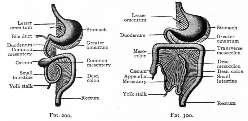
Fig. 299. Fig. 300. Diagram of the gastrointestinal tract and its mesenteries at an early and later stage
The arrow points into the bursa omentalis.
Ventral view. Hertwig.
The Greater Omentum and Omental Bursa
A small part of the gut caudal to the diaphragm is destined to become the stomach, and the portion of the mesentery which attaches it to the dorsal body wall is known as the dorsal mesogastrium (Fig. 263). The latter is inserted along the greater curvature of the stomach and lies in the medial sagittal plane so long as the stomach lies in this plane. When the stomach turns so that its long axis lies in a transverse direction and its greater curvature is directed caudally (p. 305), the dorsal mesogastrium changes its position accordingly. From its attachment along the dorsal body. wall it bends to the left and then ventrally to its attachment along the greater curvature of the stomach.
Thus a sort of sac is formed dorsal to the stomach (Figs. 299 and 300). This sac is really a part of the abdominal or peritoneal cavity and opens toward the right side. The ventral wall is formed by the stomach, the dorsal and caudal walls by the mesogastrium. The cavity of the sac is the omental bursa (bursa omentalis) ; the mesogastrium forms the greater omentum (omentum majus) . The opening from the bursa into the general peritoneal cavity is the epiploic foramen (foramen of Winslow). (Fig. 276.)
- Text-Book of Embryology: Germ cells | Maturation | Fertilization | Amphioxus | Frog | Chick | Mammalian | External body form | Connective tissues and skeletal | Vascular | Muscular | Alimentary tube and organs | Respiratory | Coelom, Diaphragm and Mesenteries | Urogenital | Integumentary | Nervous System | Special Sense | Foetal Membranes | Teratogenesis | Gallery of All Figures
| Historic Disclaimer - information about historic embryology pages |
|---|
 |
Reference
Bailey FR. and Miller AM. Text-Book of Embryology (1921) New York: William Wood and Co.
Cite this page: Hill, M.A. (2024, April 24) Embryology Bailey299 300.jpg. Retrieved from https://embryology.med.unsw.edu.au/embryology/index.php/File:Bailey299_300.jpg
- © Dr Mark Hill 2024, UNSW Embryology ISBN: 978 0 7334 2609 4 - UNSW CRICOS Provider Code No. 00098G
File history
Click on a date/time to view the file as it appeared at that time.
| Date/Time | Thumbnail | Dimensions | User | Comment | |
|---|---|---|---|---|---|
| current | 22:15, 23 January 2011 | | 911 × 447 (79 KB) | S8600021 (talk | contribs) | {{Template:Bailey 1921 Figures}} Category:Human Category:Coelom |
You cannot overwrite this file.
File usage
The following 5 pages use this file: