File:Bailey278 279.jpg

Original file (669 × 812 pixels, file size: 85 KB, MIME type: image/jpeg)
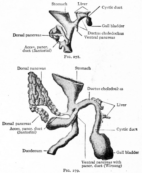
Figs. 278 and 279. From models of the developing liver and pancreas of rabbit embryos
Rabbit embryos of 8 mm. and 10 mm. respectively
Both seen from the right side.
Hammar, Bonnet.
The Development of the Pancreas
The epithelium of the pancreas, like that of the liver, is a derivative of the entoderm. It arises from two (or three) separate anlagen, one dorsal and one (or two) ventral. The dorsal anlage appears first as a ridge-like evagination from the dorsal wall of the gut, slightly cranial to the level of the liver (Figs. 273 and 274). It appears about the same time as the liver or a little later. The mass of cells grows into the dorsal mesentery and becomes constricted from the parent epithelium except for a thin neck which becomes the duct of Santorini (Fig. 278). A little later two other diverticula appear, one from each side of the common bile duct. It is uncertain whether only one or both of these take part in the formation of the pancreas, but it seems most probable that the left one disappears entirely. The right diverticulum continues to develop and becomes constricted from the parent epithelium, leaving only a thin neck which becomes the duct of Wirsung.
The smaller ventral pancreas grows to the right and then dorsally in the mesentery (Fig. 260), passing over the right surface of the portal vein, until it meets and fuses with the proximal part of the larger dorsal pancreas. The fusion takes place in the sixth week, and the two anlagen then form a single mass. A communication is established between the two ducts, and the dorsal duct (Santorini) usually disappears, leaving the ventral (Wirsung) as the permanent duct opening into the ductus choledochus. In a general way it may be said that the ventral anlage gives rise to the head, the dorsal anlage to the body and tail of the pancreas (compare Figs. 278 and 279).
As the pancreas grows into the dorsal mesentery it comes to lie in the dorsal mesogastrium between the greater curvature of the stomach and the vertebral column, and since the dorsal mesogastrium at first lies in the medial sagittal plane, the pancreas is similarly situated. After the sixth week, however, as the stomach changes its position (p. 305) , the pancreas is carried along with the mesogastrium and comes to lie in a transverse plane, with its head to the right and embedded in the bend of the duodenum, and its tail reaching to the spleen on the left. The organ as a whole is at first movable along with the mesentery, but when it assumes its transverse position it lies close to the dorsal abdominal wall. The mesentery then fuses with the adjacent peritoneum (see p. 350), and the pancreas is firmly fixed.
- Text-Book of Embryology: Germ cells | Maturation | Fertilization | Amphioxus | Frog | Chick | Mammalian | External body form | Connective tissues and skeletal | Vascular | Muscular | Alimentary tube and organs | Respiratory | Coelom, Diaphragm and Mesenteries | Urogenital | Integumentary | Nervous System | Special Sense | Foetal Membranes | Teratogenesis | Gallery of All Figures
| Historic Disclaimer - information about historic embryology pages |
|---|
 |
Reference
Bailey FR. and Miller AM. Text-Book of Embryology (1921) New York: William Wood and Co.
Cite this page: Hill, M.A. (2024, April 19) Embryology Bailey278 279.jpg. Retrieved from https://embryology.med.unsw.edu.au/embryology/index.php/File:Bailey278_279.jpg
- © Dr Mark Hill 2024, UNSW Embryology ISBN: 978 0 7334 2609 4 - UNSW CRICOS Provider Code No. 00098G
File history
Click on a date/time to view the file as it appeared at that time.
| Date/Time | Thumbnail | Dimensions | User | Comment | |
|---|---|---|---|---|---|
| current | 22:04, 23 January 2011 | | 669 × 812 (85 KB) | S8600021 (talk | contribs) | {{Template:Bailey 1921 Figures}} Category:Human Category:Gastrointestinal Tract Category:Liver |
You cannot overwrite this file.
File usage
The following 4 pages use this file: