File:Bailey274.jpg
Bailey274.jpg (558 × 442 pixels, file size: 32 KB, MIME type: image/jpeg)
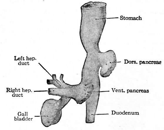
Fig. 274. From a reconstruction of the anlagen of the liver and pancreas and a part of the stomach and duodenum of a human embryo of 4 weeks
Felix.
The liver is the first gland of the digestive tract to appear. In embryos of about 3 mm. a longitudinal ridge-like evagination develops from the entoderm on the ventral side of the gut a short distance caudal to the stomach, that is, in the duodenal portion of the gut (Figs. 247, 272, 273). The cephalic part of the evagination is solid and, being destined to give rise to the liver proper, is called the pars hepatica. The caudal part is hollow, its cavity being continuous with the lumen of the gut, and is destined to give rise to the gall bladder, whence it is called the pars cystica. Beginning at both the cephalic and caudal ends, the evagination as a whole becomes constricted from the gut until (in embryos of about 8 mm.) its only connection with the latter is a narrow cord of cells which is the anlage of the ductus choledochus. The pars hepatica by this time has enlarged considerably and remains attached to the ductus choledochus by a short cord of cells, the anlage of the hepatic duct. The pars cystica has also become larger, its distal portion being somewhat dilated, and is connected with the ductus choledochus by the anlage of the cystic duct (Figs. 274 and 275). The pars cystica grows into the ventral mesentery and thus becomes surrounded by mesodermal tissue. The proximal portion continues to elongate to form the cystic duct and the distal portion becomes larger and more dilated to form the gall bladder.
- Text-Book of Embryology: Germ cells | Maturation | Fertilization | Amphioxus | Frog | Chick | Mammalian | External body form | Connective tissues and skeletal | Vascular | Muscular | Alimentary tube and organs | Respiratory | Coelom, Diaphragm and Mesenteries | Urogenital | Integumentary | Nervous System | Special Sense | Foetal Membranes | Teratogenesis | Gallery of All Figures
| Historic Disclaimer - information about historic embryology pages |
|---|
 |
Reference
Bailey FR. and Miller AM. Text-Book of Embryology (1921) New York: William Wood and Co.
Cite this page: Hill, M.A. (2024, April 19) Embryology Bailey274.jpg. Retrieved from https://embryology.med.unsw.edu.au/embryology/index.php/File:Bailey274.jpg
- © Dr Mark Hill 2024, UNSW Embryology ISBN: 978 0 7334 2609 4 - UNSW CRICOS Provider Code No. 00098G
File history
Click on a date/time to view the file as it appeared at that time.
| Date/Time | Thumbnail | Dimensions | User | Comment | |
|---|---|---|---|---|---|
| current | 22:02, 23 January 2011 | | 558 × 442 (32 KB) | S8600021 (talk | contribs) | {{Template:Bailey 1921 Figures}} Category:Human Category:Gastrointestinal Tract Category:Liver |
You cannot overwrite this file.