File:Bailey081.jpg
Original file (782 × 755 pixels, file size: 118 KB, MIME type: image/jpeg)
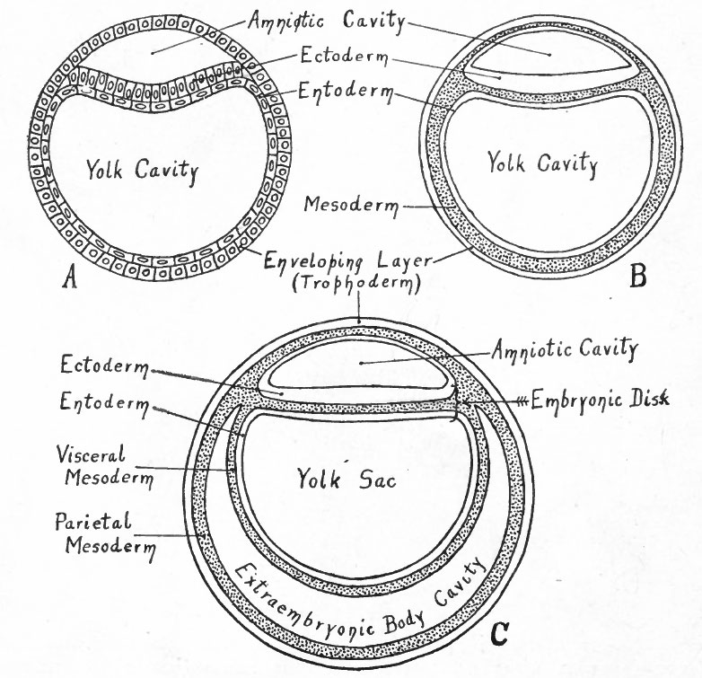
Fig. 81. Diagrams representing hypothetical stages in the development of the human embryo
(to follow Fig. 80).
A, Entoderm surrounds the yolk cavity; part of the cells of the inner cell mass have become vacuolated, thus forming the amniotic cavity, while the remainder constitute the embryonic ectoderm; compare with Fig. 59.
B, Mesoderm (represented by dotted portion) has appeared between the entoderm and trophoderm, between the entoderm and ectoderm of the embryonic disk, and in the roof of the amnion.
C, The mesoderm around the yolk cavity has split into a parietal and a visceral layer, the cleft between being the rudiment of the extraembryonic body cavity (exoccelom).
The series of diagrams in Figs. 80, 81 and 82 has been constructed to give the student a general idea of the changes that occur in the early stages of human development. It must be recognized, however, that the diagrams represent purely hypothetical stages up to the conditions shown in diagram B in Fig. 81 which corresponds roughly to the Bryce-Teacher embryo (Fig. 73) ; even in this diagram the extent of the mesoderm is much less than in the known human embryo. In Fig. 82 diagram A approximates the Peters embryo (Fig. 74), diagram D the von Spee embryo (Fig. 77). The history of the accessory structures which are shown in part will be considered in the chapter on "Foetal Membranes".
- Text-Book of Embryology: Germ cells | Maturation | Fertilization | Amphioxus | Frog | Chick | Mammalian | External body form | Connective tissues and skeletal | Vascular | Muscular | Alimentary tube and organs | Respiratory | Coelom, Diaphragm and Mesenteries | Urogenital | Integumentary | Nervous System | Special Sense | Foetal Membranes | Teratogenesis | Gallery of All Figures
| Historic Disclaimer - information about historic embryology pages |
|---|
 |
Reference
Bailey FR. and Miller AM. Text-Book of Embryology (1921) New York: William Wood and Co.
Cite this page: Hill, M.A. (2024, April 18) Embryology Bailey081.jpg. Retrieved from https://embryology.med.unsw.edu.au/embryology/index.php/File:Bailey081.jpg
- © Dr Mark Hill 2024, UNSW Embryology ISBN: 978 0 7334 2609 4 - UNSW CRICOS Provider Code No. 00098G
File history
Click on a date/time to view the file as it appeared at that time.
| Date/Time | Thumbnail | Dimensions | User | Comment | |
|---|---|---|---|---|---|
| current | 11:28, 18 January 2011 | | 782 × 755 (118 KB) | S8600021 (talk | contribs) | |
| 11:24, 18 January 2011 |  | 1,630 × 1,200 (289 KB) | S8600021 (talk | contribs) | {{Template:Bailey 1921 Figures}} |
You cannot overwrite this file.
File usage
The following 2 pages use this file: