File:Bailey073.jpg: Difference between revisions
From Embryology
No edit summary |
No edit summary |
||
| (2 intermediate revisions by the same user not shown) | |||
| Line 12: | Line 12: | ||
* P.e., point of entrance of the ovum | * P.e., point of entrance of the ovum | ||
* tro., syncytial layer (plasmodi-trophoderm) | * tro., syncytial layer (plasmodi-trophoderm) | ||
* tro. 1 , masses of vacuolating syncytium invading capillaries. | * tro.<sup>1</sup> , masses of vacuolating syncytium invading capillaries. | ||
The cavity of the vesicle is filled with mesoderm in which are embedded the amniotic cavity (the larger) and the yolk cavity. | The cavity of the vesicle is filled with mesoderm in which are embedded the amniotic cavity (the larger) and the yolk cavity. | ||
:"In certain respects the Bryce-Teacher embryo (Fig. 73) bears fundamental resemblances to corresponding stages of lower mammals, especially the lower primates; in other respects there are differences which are not irreconcilable, however, with the general principles of mammalian ontogeny. The vesiclelike structure of the entire developing organism is a fairly close approximation to the trophodermal sac of the lower forms. In both cases the rudiment of the embryonic body is contained within the sac. In the human embryo in question there are two cavities within the vesicle; the larger is regarded as the amniotic cavity lined with ectoderm, and the smaller as the cavity of the yolk sac lined with entoderm. The double wall between the two would be the embryonic disk. The precocious development of the mesoderm, which as a loosely arranged tissue fills in all the space between the trophoderm and the two small cavities, is one of the remarkable features of this embryo. The trophoderm is a most elaborate layer and has sent out irregular projections into the uterine mucosa in which the whole structure is already embedded. The early embedding or implantation and the elaboration of the trophoderm are probably closely correlated." | |||
{{Template:Bailey 1921 Figures}} | {{Template:Bailey 1921 Figures}} | ||
[[Category:Human]] | [[Category:Coelomic Cavity]] [[Category:Human]] [[Category:Uterus]] [[Category:Implantation]] [[Category:Week 2]] | ||
Latest revision as of 04:08, 13 April 2011
Fig. 73. Section of a human ovum of about 14 days, embedded in the uterine mucosa
Bryce and Teacher.
Legend
- Cap., Capillary
- cyt., cellular layer (cyto-trophoderm)
- ep., uterine epithelium
- gl., uterine gland
- n.z., necrotic zone of decidua (uterine mucosa)
- P.e., point of entrance of the ovum
- tro., syncytial layer (plasmodi-trophoderm)
- tro.1 , masses of vacuolating syncytium invading capillaries.
The cavity of the vesicle is filled with mesoderm in which are embedded the amniotic cavity (the larger) and the yolk cavity.
- "In certain respects the Bryce-Teacher embryo (Fig. 73) bears fundamental resemblances to corresponding stages of lower mammals, especially the lower primates; in other respects there are differences which are not irreconcilable, however, with the general principles of mammalian ontogeny. The vesiclelike structure of the entire developing organism is a fairly close approximation to the trophodermal sac of the lower forms. In both cases the rudiment of the embryonic body is contained within the sac. In the human embryo in question there are two cavities within the vesicle; the larger is regarded as the amniotic cavity lined with ectoderm, and the smaller as the cavity of the yolk sac lined with entoderm. The double wall between the two would be the embryonic disk. The precocious development of the mesoderm, which as a loosely arranged tissue fills in all the space between the trophoderm and the two small cavities, is one of the remarkable features of this embryo. The trophoderm is a most elaborate layer and has sent out irregular projections into the uterine mucosa in which the whole structure is already embedded. The early embedding or implantation and the elaboration of the trophoderm are probably closely correlated."
- Text-Book of Embryology: Germ cells | Maturation | Fertilization | Amphioxus | Frog | Chick | Mammalian | External body form | Connective tissues and skeletal | Vascular | Muscular | Alimentary tube and organs | Respiratory | Coelom, Diaphragm and Mesenteries | Urogenital | Integumentary | Nervous System | Special Sense | Foetal Membranes | Teratogenesis | Gallery of All Figures
| Historic Disclaimer - information about historic embryology pages |
|---|
 |
Reference
Bailey FR. and Miller AM. Text-Book of Embryology (1921) New York: William Wood and Co.
Cite this page: Hill, M.A. (2024, April 16) Embryology Bailey073.jpg. Retrieved from https://embryology.med.unsw.edu.au/embryology/index.php/File:Bailey073.jpg
- © Dr Mark Hill 2024, UNSW Embryology ISBN: 978 0 7334 2609 4 - UNSW CRICOS Provider Code No. 00098G
File history
Click on a date/time to view the file as it appeared at that time.
| Date/Time | Thumbnail | Dimensions | User | Comment | |
|---|---|---|---|---|---|
| current | 17:01, 17 January 2011 | 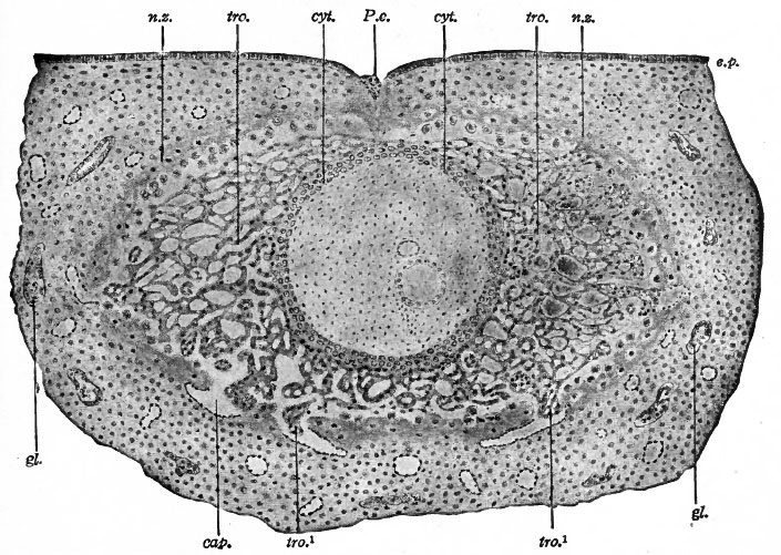 | 705 × 501 (122 KB) | S8600021 (talk | contribs) | {{Template:Bailey 1921 Figures}} |
You cannot overwrite this file.
File usage
The following 2 pages use this file: