File:Bailey007.jpg: Difference between revisions
({{Template:Bailey 1921 Figures}}) |
No edit summary |
||
| Line 1: | Line 1: | ||
==Fig. 7. Reduction of chromosomes in spermatogenesis in Ascaris megalocephala (bivalens)== | |||
Brauer, Wilson. | |||
A - G Successive stages in the division of the primary spermatocyte. | |||
The original reticulum undergoes a very early division of the chromatin granules which then form a doubly split spireme (B). This becomes shorter (C), and then breaks in two to form the 2 tetrads (D, in profile, E, on end). F, G, H, First division to form 2 secondary spermatocytes, each receiving 2 dyads. I, Secondary spermatocyte. J, K, The same dividing. L, Two resulting spermatids, each containing 2 single chromosomes. | |||
{{Template:Bailey 1921 Figures}} | {{Template:Bailey 1921 Figures}} | ||
[[Category:Spermatozoa]] | |||
Latest revision as of 13:49, 12 February 2011
Fig. 7. Reduction of chromosomes in spermatogenesis in Ascaris megalocephala (bivalens)
Brauer, Wilson.
A - G Successive stages in the division of the primary spermatocyte.
The original reticulum undergoes a very early division of the chromatin granules which then form a doubly split spireme (B). This becomes shorter (C), and then breaks in two to form the 2 tetrads (D, in profile, E, on end). F, G, H, First division to form 2 secondary spermatocytes, each receiving 2 dyads. I, Secondary spermatocyte. J, K, The same dividing. L, Two resulting spermatids, each containing 2 single chromosomes.
- Text-Book of Embryology: Germ cells | Maturation | Fertilization | Amphioxus | Frog | Chick | Mammalian | External body form | Connective tissues and skeletal | Vascular | Muscular | Alimentary tube and organs | Respiratory | Coelom, Diaphragm and Mesenteries | Urogenital | Integumentary | Nervous System | Special Sense | Foetal Membranes | Teratogenesis | Gallery of All Figures
| Historic Disclaimer - information about historic embryology pages |
|---|
 |
Reference
Bailey FR. and Miller AM. Text-Book of Embryology (1921) New York: William Wood and Co.
Cite this page: Hill, M.A. (2024, April 18) Embryology Bailey007.jpg. Retrieved from https://embryology.med.unsw.edu.au/embryology/index.php/File:Bailey007.jpg
- © Dr Mark Hill 2024, UNSW Embryology ISBN: 978 0 7334 2609 4 - UNSW CRICOS Provider Code No. 00098G
File history
Click on a date/time to view the file as it appeared at that time.
| Date/Time | Thumbnail | Dimensions | User | Comment | |
|---|---|---|---|---|---|
| current | 14:35, 17 January 2011 | 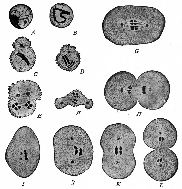 | 772 × 803 (138 KB) | S8600021 (talk | contribs) | {{Template:Bailey 1921 Figures}} |
You cannot overwrite this file.
File usage
The following 2 pages use this file: