File:Atwell1918 fig39.jpg
From Embryology

Size of this preview: 507 × 599 pixels. Other resolution: 600 × 709 pixels.
Original file (600 × 709 pixels, file size: 139 KB, MIME type: image/jpeg)
Fig. 39 Another detail drawing of the pars tuberalis. X 400. b.w., brain wall; p.t., pars tuberalis. Several bloodvessels are shown in the pia mater.
File history
Click on a date/time to view the file as it appeared at that time.
| Date/Time | Thumbnail | Dimensions | User | Comment | |
|---|---|---|---|---|---|
| current | 11:08, 10 November 2016 | | 600 × 709 (139 KB) | Z8600021 (talk | contribs) | |
| 11:07, 10 November 2016 | 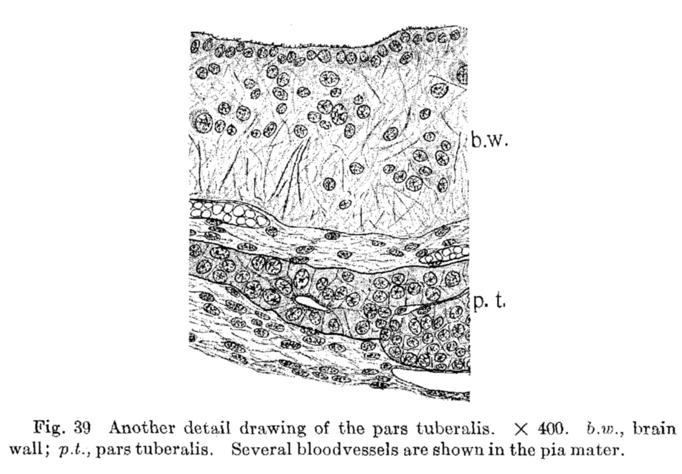 | 1,333 × 927 (185 KB) | Z8600021 (talk | contribs) | Fig. 39 Another detail drawing of the pars tuberalis. X 400. b.w., brain wall; p.t., pars tuberalis. Several bloodvessels are shown in the pia mater. |
You cannot overwrite this file.
File usage
The following page uses this file: