File:Atwell1918 fig22.jpg: Difference between revisions
No edit summary |
mNo edit summary |
||
| (One intermediate revision by the same user not shown) | |||
| Line 1: | Line 1: | ||
==Fig. 22 Model of hypophysis region, seventeen-day embryo== | |||
(28A), viewed from right side. X 50. b.w., brain waII; n.Z., neural lobe; p.t., pars tuberalis; cort., cortical plate; sphen., portion of sphenoid; st., stalk; p . , process surrounding neck of neural lobe. | |||
{{Historic Disclaimer}} | |||
See also {{Ref-Atwell1926}} | |||
'''Links:''' [[Endocrine - Pituitary Development|Pituitary Development]] | [[Rabbit Development]] | |||
===Reference=== | |||
{{Ref-Atwell1918}} | |||
{{Footer}} | |||
[[Category:Pituitary]][[Category:Rabbit]][[Category:1910's]] | |||
Latest revision as of 18:36, 14 November 2016
Fig. 22 Model of hypophysis region, seventeen-day embryo
(28A), viewed from right side. X 50. b.w., brain waII; n.Z., neural lobe; p.t., pars tuberalis; cort., cortical plate; sphen., portion of sphenoid; st., stalk; p . , process surrounding neck of neural lobe.
| Historic Disclaimer - information about historic embryology pages |
|---|
 |
See also Atwell WJ. The development of the hypophysis cerebri in man, with special reference to the pars tuberalis. (1926) Amer. J Anat. 37: 139-193.
Links: Pituitary Development | Rabbit Development
Reference
Atwell WJ. The development of the hypophysis cerebri of the rabbit (Lepus Cuniculus L.). (1918) Amer. J Anat. 24(2): 271-337
Cite this page: Hill, M.A. (2024, April 16) Embryology Atwell1918 fig22.jpg. Retrieved from https://embryology.med.unsw.edu.au/embryology/index.php/File:Atwell1918_fig22.jpg
- © Dr Mark Hill 2024, UNSW Embryology ISBN: 978 0 7334 2609 4 - UNSW CRICOS Provider Code No. 00098G
File history
Click on a date/time to view the file as it appeared at that time.
| Date/Time | Thumbnail | Dimensions | User | Comment | |
|---|---|---|---|---|---|
| current | 18:36, 14 November 2016 | 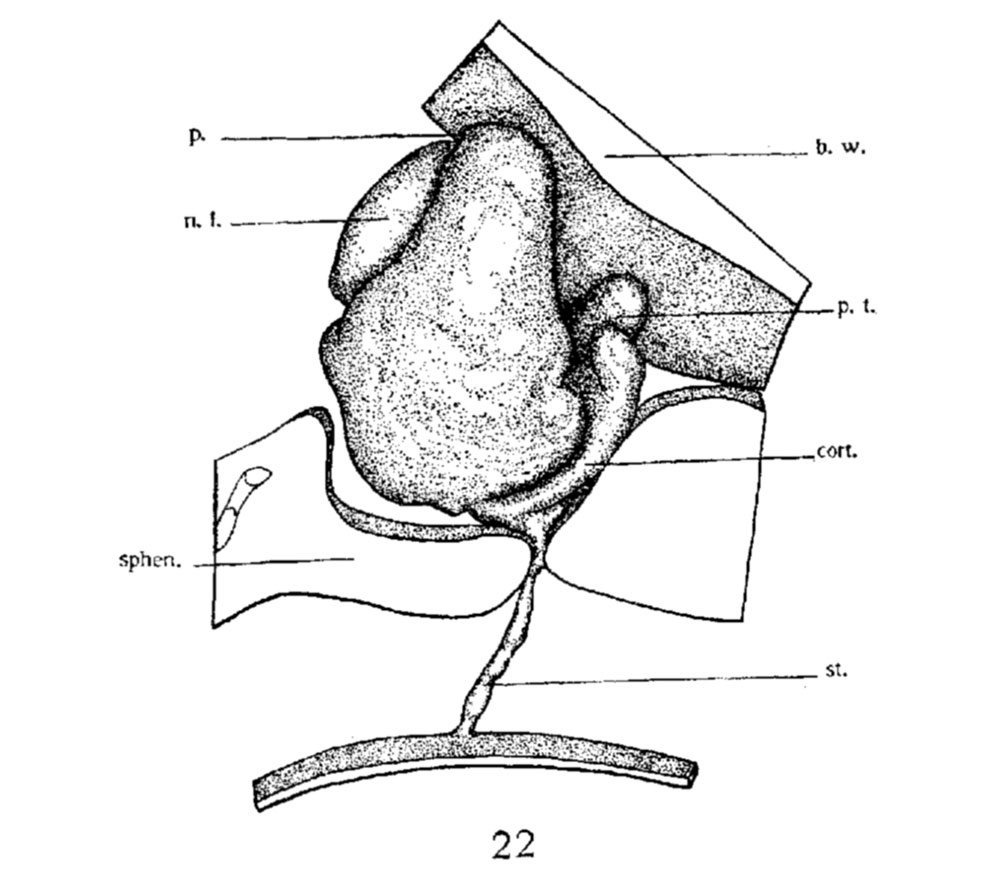 | 1,000 × 887 (100 KB) | Z8600021 (talk | contribs) | |
| 18:35, 14 November 2016 | 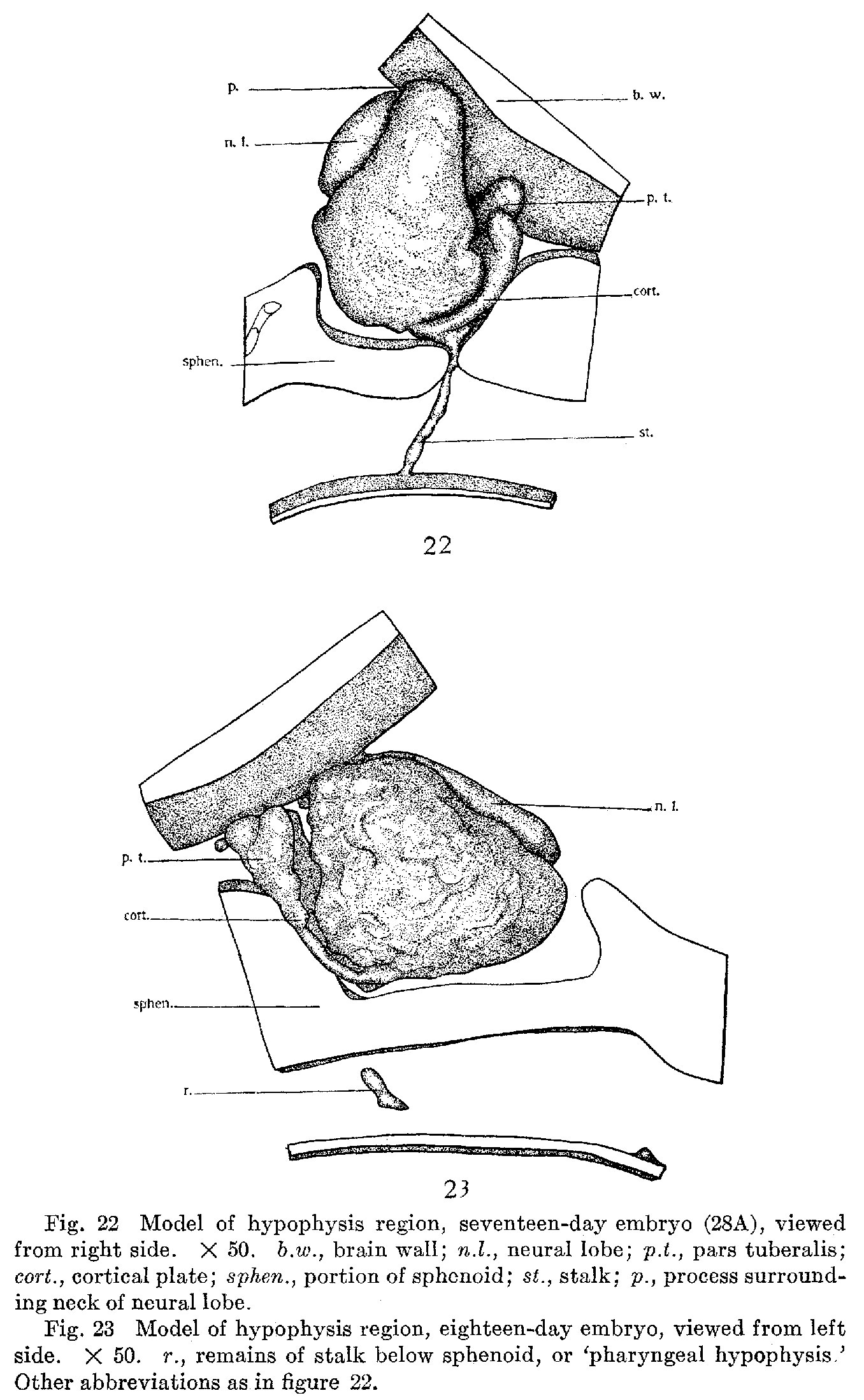 | 1,356 × 2,198 (465 KB) | Z8600021 (talk | contribs) |
You cannot overwrite this file.
File usage
The following page uses this file: