File:Arey1924 fig418.jpg: Difference between revisions
(Z8600021 uploaded a new version of File:Arey1924 fig418.jpg) |
mNo edit summary |
||
| (One intermediate revision by the same user not shown) | |||
| Line 1: | Line 1: | ||
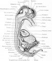
==Fig. 418. Median sagittal dissection of a 35 mm Embryo== | |||
original x4 | |||
Median Sagittal Section (Fig. 418). - New features of the brain are the olfactory lobes, the chorioid plexus of the third and lateral ventricles, the thalami, the epiphysis, and consolidated hypophysis. The primitive mouth cavity is now divided by the palatine folds into upper nasal passages and lower oral cavity. Of the viscera, the distinct genital and suprarenal glands and the enlarged metanephros command attention, as does the coiling of the intestine. The ureter has acquired a separate opening at the base of the bladder, and the urethra may be traced into the phallus. | |||
{{Arey1924 Footer}} | |||
[[Category:Pig]] | |||
Latest revision as of 17:20, 4 January 2017
Fig. 418. Median sagittal dissection of a 35 mm Embryo
original x4
Median Sagittal Section (Fig. 418). - New features of the brain are the olfactory lobes, the chorioid plexus of the third and lateral ventricles, the thalami, the epiphysis, and consolidated hypophysis. The primitive mouth cavity is now divided by the palatine folds into upper nasal passages and lower oral cavity. Of the viscera, the distinct genital and suprarenal glands and the enlarged metanephros command attention, as does the coiling of the intestine. The ureter has acquired a separate opening at the base of the bladder, and the urethra may be traced into the phallus.
| Historic Disclaimer - information about historic embryology pages |
|---|
| Pages where the terms "Historic" (textbooks, papers, people, recommendations) appear on this site, and sections within pages where this disclaimer appears, indicate that the content and scientific understanding are specific to the time of publication. This means that while some scientific descriptions are still accurate, the terminology and interpretation of the developmental mechanisms reflect the understanding at the time of original publication and those of the preceding periods, these terms, interpretations and recommendations may not reflect our current scientific understanding. (More? Embryology History | Historic Embryology Papers) |
Reference
Arey LB. Developmental Anatomy. (1924) W.B. Saunders Company, Philadelphia.
Cite this page: Hill, M.A. (2024, April 18) Embryology Arey1924 fig418.jpg. Retrieved from https://embryology.med.unsw.edu.au/embryology/index.php/File:Arey1924_fig418.jpg
- © Dr Mark Hill 2024, UNSW Embryology ISBN: 978 0 7334 2609 4 - UNSW CRICOS Provider Code No. 00098G
File history
Click on a date/time to view the file as it appeared at that time.
| Date/Time | Thumbnail | Dimensions | User | Comment | |
|---|---|---|---|---|---|
| current | 17:18, 4 January 2017 |  | 1,280 × 1,498 (343 KB) | Z8600021 (talk | contribs) | |
| 17:17, 4 January 2017 |  | 1,280 × 1,553 (353 KB) | Z8600021 (talk | contribs) |
You cannot overwrite this file.
File usage
The following 2 pages use this file:

