File:Arey1924 fig195.jpg
Original file (1,583 × 950 pixels, file size: 267 KB, MIME type: image/jpeg)
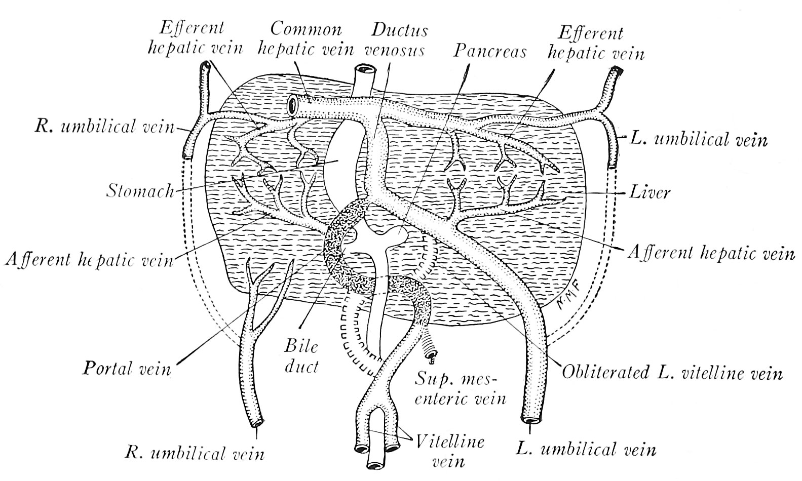
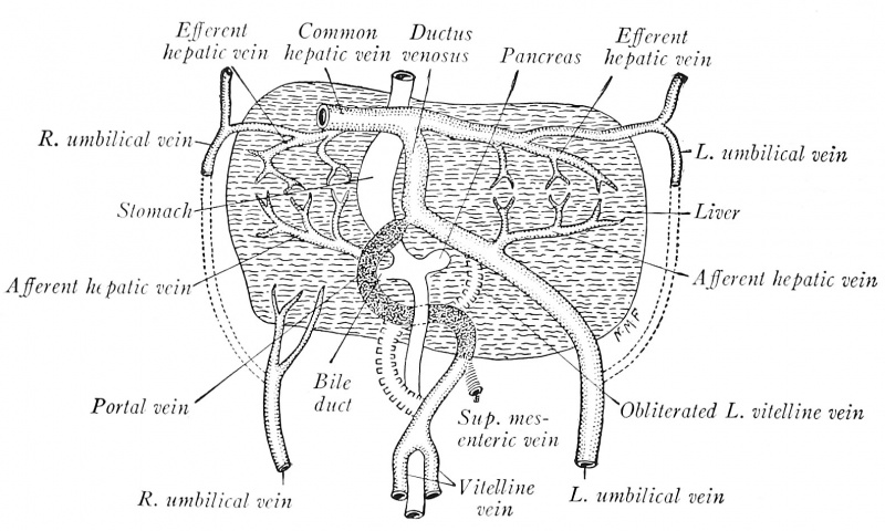
Fig. 195. The origin of the portal vein and ductus venosus as illustrated by a human embryo of 7 mm
modified after Wilhelm His (1831-1904).
In the liver, the portal vein, through its cranial anastomosis between the primitive vitelline veins, is connected with the left umbilical vein (Fig. 195). As the right lobe of the liver grows, the course of the umbilical and portal blood through the intrahepatic portion of the right vitelline vein becomes circuitous, and hence a new, direct channel to the sinus venosus is formed through the hepatic sinusoids. This is the ductus venosus (Fig. 195), which is obliterated after birth and forms the ligamentum venosum of the postnatal liver.
| Historic Disclaimer - information about historic embryology pages |
|---|
 |

Reference
Arey LB. Developmental Anatomy. (1924) W.B. Saunders Company, Philadelphia.
Cite this page: Hill, M.A. (2024, April 18) Embryology Arey1924 fig195.jpg. Retrieved from https://embryology.med.unsw.edu.au/embryology/index.php/File:Arey1924_fig195.jpg
- © Dr Mark Hill 2024, UNSW Embryology ISBN: 978 0 7334 2609 4 - UNSW CRICOS Provider Code No. 00098G
File history
Click on a date/time to view the file as it appeared at that time.
| Date/Time | Thumbnail | Dimensions | User | Comment | |
|---|---|---|---|---|---|
| current | 14:04, 23 October 2016 | | 1,583 × 950 (267 KB) | Z8600021 (talk | contribs) | |
| 14:02, 23 October 2016 | 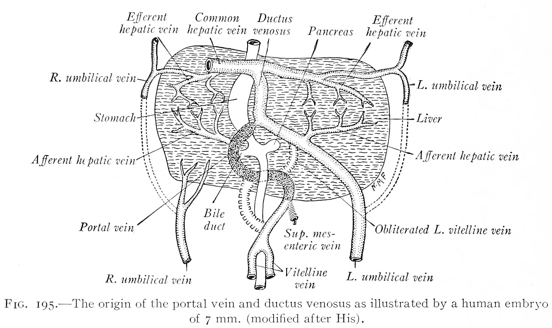 | 1,767 × 1,050 (297 KB) | Z8600021 (talk | contribs) | ==Fig. 195. The origin of the portal vein and ductus venosus as illustrated by a human embryo of 7 mm== (modified after His). |
You cannot overwrite this file.
File usage
The following 2 pages use this file: