File:Arey1924 fig195.jpg: Difference between revisions
From Embryology
(==Fig. 195. The origin of the portal vein and ductus venosus as illustrated by a human embryo of 7 mm== (modified after His).) |
mNo edit summary |
||
| Line 1: | Line 1: | ||
==Fig. 195. The origin of the portal vein and ductus venosus as illustrated by a human embryo of 7 mm== | ==Fig. 195. The origin of the portal vein and ductus venosus as illustrated by a human embryo of 7 mm== | ||
(modified after His). | (modified after {{His}}). | ||
{{Arey1924 Footer}} | |||
[[Category:Cardiovascular]] | |||
Revision as of 14:03, 23 October 2016
Fig. 195. The origin of the portal vein and ductus venosus as illustrated by a human embryo of 7 mm
(modified after Wilhelm His (1831-1904)).
| Historic Disclaimer - information about historic embryology pages |
|---|
 |

Reference
Arey LB. Developmental Anatomy. (1924) W.B. Saunders Company, Philadelphia.
Cite this page: Hill, M.A. (2024, April 25) Embryology Arey1924 fig195.jpg. Retrieved from https://embryology.med.unsw.edu.au/embryology/index.php/File:Arey1924_fig195.jpg
- © Dr Mark Hill 2024, UNSW Embryology ISBN: 978 0 7334 2609 4 - UNSW CRICOS Provider Code No. 00098G
File history
Click on a date/time to view the file as it appeared at that time.
| Date/Time | Thumbnail | Dimensions | User | Comment | |
|---|---|---|---|---|---|
| current | 14:04, 23 October 2016 | 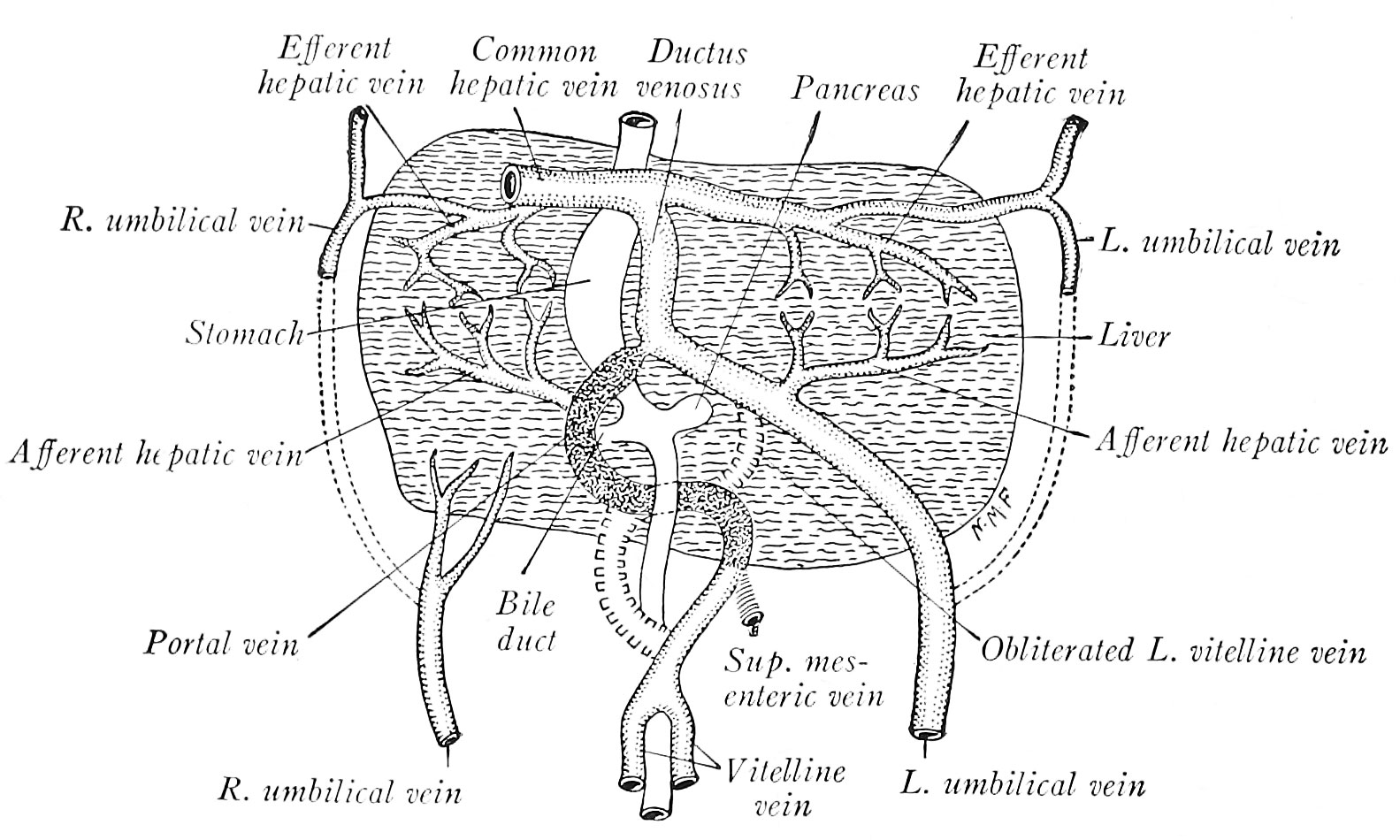 | 1,583 × 950 (267 KB) | Z8600021 (talk | contribs) | |
| 14:02, 23 October 2016 | 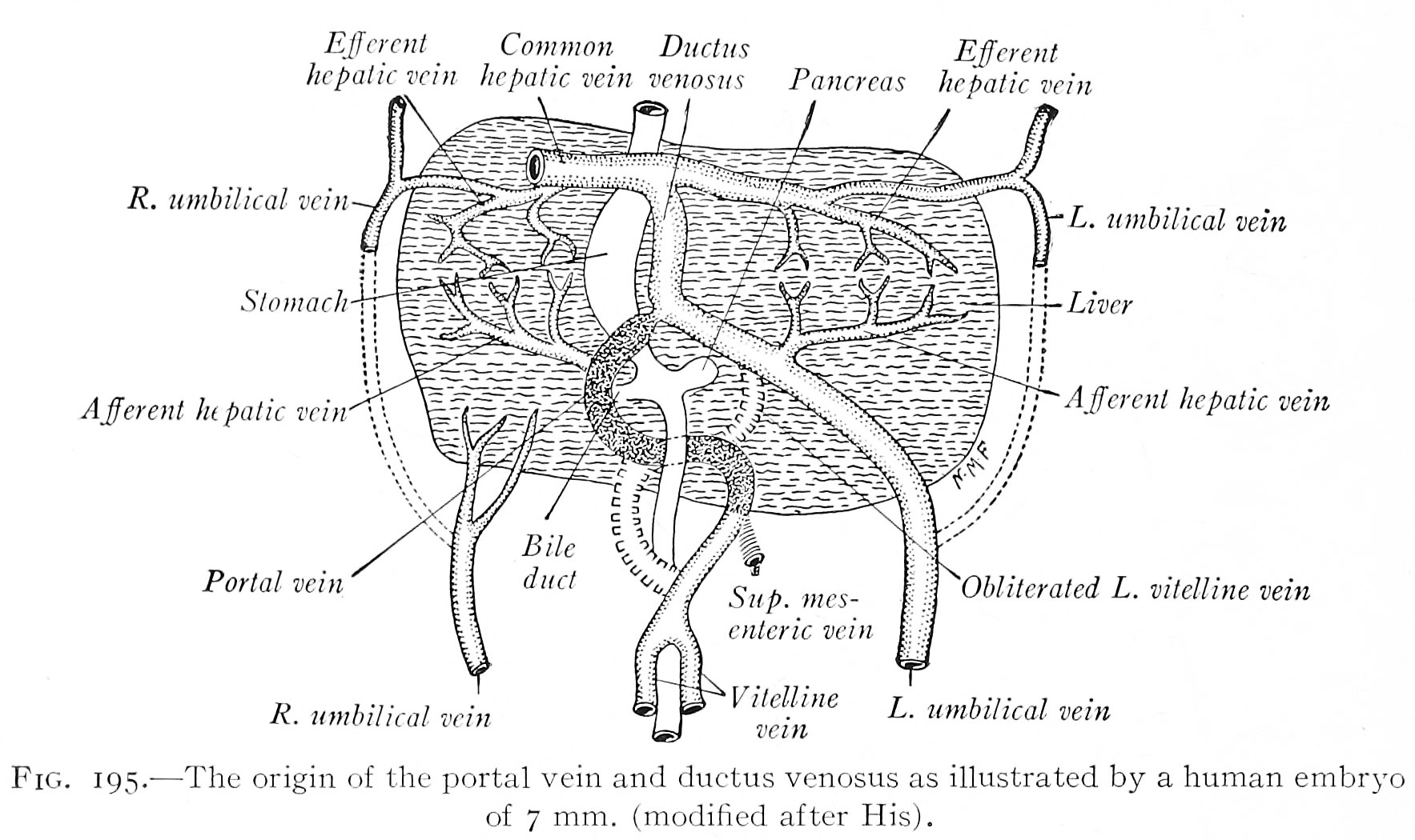 | 1,767 × 1,050 (297 KB) | Z8600021 (talk | contribs) | ==Fig. 195. The origin of the portal vein and ductus venosus as illustrated by a human embryo of 7 mm== (modified after His). |
You cannot overwrite this file.
File usage
The following 2 pages use this file: