File:Anson1948 fig16.jpg

Original file (1,280 × 817 pixels, file size: 133 KB, MIME type: image/jpeg)
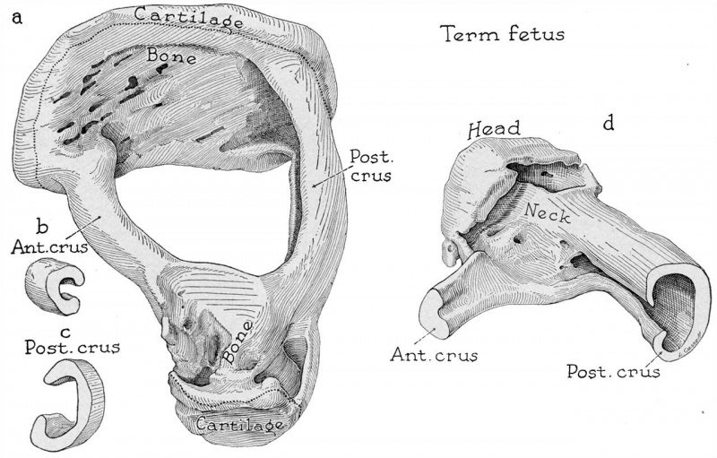
Fig. 16. Drawings of a reconstruction of the stapes in a fetus at term
(Wisconsin series 102) ; x 7: (a) the reconstruction entire, seen from a superolateral position; (b) segment of anterior crus; (c) segment of posterior crus; (d) lateral part of the reconstruction, in posteromedial view. In a are indicated the limits of the segments shown in b and c.
The cartilage which covers the vestibular (medial) surface of the base is carried over the fenestral margin as a circumferential lip, while the tympanic (lateral) surface is formed by a plate of irregular bone which is composed of both endochondral and perichondrial bone (:2; cf. figs. 13a and 14 a). The crura are deeply channeled (16a to c). The head and neck of the ossicle are strikingly eroded, despite the fact that the cavity of the capital portion is crossed by a plate of bone (a and d). This feature of sculpturing is persistent, being encountered in ossicles from adults (specimens from a 57 year old person and from other adults). The anterior crus, much the slenderer and shorter of the crura, is implanted in the base at the inferior margin of the crus (a); at the point of continuity with the head, the crus flattens to meet the intercrural plate (d). The bulkier posterior crus is implanted widely into its portion of the base (a); its cavity, relatively capacious (c), opens into that of the head and neck by a small orifice (d). Cartilage covers the articular surface of the head as a layer of restricted extent (a).
Reference
Anson BJ. and Cauldwell EW. Stapes, fissula ante fenestram and associated structures in man: V . From the fetus of 160 mm to term. (1948) 48(3): 263-300.
Cite this page: Hill, M.A. (2024, April 25) Embryology Anson1948 fig16.jpg. Retrieved from https://embryology.med.unsw.edu.au/embryology/index.php/File:Anson1948_fig16.jpg
- © Dr Mark Hill 2024, UNSW Embryology ISBN: 978 0 7334 2609 4 - UNSW CRICOS Provider Code No. 00098G
File history
Click on a date/time to view the file as it appeared at that time.
| Date/Time | Thumbnail | Dimensions | User | Comment | |
|---|---|---|---|---|---|
| current | 15:55, 13 October 2017 | | 1,280 × 817 (133 KB) | Z8600021 (talk | contribs) | |
| 15:47, 13 October 2017 |  | 1,559 × 1,672 (374 KB) | Z8600021 (talk | contribs) | ==Fig. 16. == ===Reference=== {{Ref-Anson1948}} {{Footer}} |
You cannot overwrite this file.
File usage
The following 2 pages use this file: