File:Anson1948 fig08.jpg

Original file (1,280 × 1,764 pixels, file size: 207 KB, MIME type: image/jpeg)
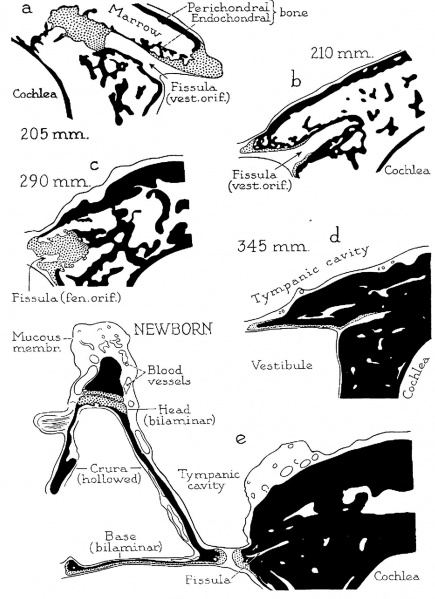
Fig. 8. Otic capsule developmental stages 205, 210, 290, 345, 345 mm Fetus and Newborn Infant
Drawings of developmental stages concluded. X 6.6. (a) 205 mm. (23 week) fetus (Wisconsin series 129, slide 20, section 3); (b) 210 mm. (23 week) fetus (Wisconsin series 51, slide 38, section 5); (C) 290 mm. (32 week) fetus (Wisconsi11 series 59, slide 36, section 5); (d) 345 mm. (38 week), fetus (Wisconsin series 61, slide 42, section 1); (e) newborn infant (Wisconsin series 315. slide 20', section 8). Parts a, b and d are from sections through the vestibular orifice of the fissula; (C) and (e), from sections at the fenestral orifice.
The cartilage which surrounds the fissula ante fenestram in the 202 mm. specimen (fig. 6b) as a thick chondral layer, bordered by spicules of intrachondrial bone, undergoes rapid reduction in bulk to become, at the vestibular extremity, an osseous shell lined by a thin stratum of persistent cartilage or by a perichondrium (205 and 210 mm., a and b). Cartilage still persists at the cochlear extremity of the fissula as a considerable mass (205 mm., a); similarly, it still remains at the fenestral extremity, where the fissular cartilage is still continuous with similar tissue which lines the vestibular window (290 mm., c). While the cartilage is being gradually reduced in bulk, the marrow space, which everywhere surrounds the fissula, becomes occupied by endochondral bone. At first, spicules of_intrachondrial bone are sparsely distributed in the area between plates of perichondrial bone (205 and 210 mm., a and b); endochondral bone next forms around these spicules (290 mm., C), finally converting the capsule into a petrous structure (345 mm., d; newborn, 9). Thus, in the newborn (c), the capsule has acquired an adult appearance, as has the stapes.
Reference
Anson BJ. and Cauldwell EW. Stapes, fissula ante fenestram and associated structures in man: V . From the fetus of 160 mm to term. (1948) 48(3): 263-300.
Cite this page: Hill, M.A. (2024, April 25) Embryology Anson1948 fig08.jpg. Retrieved from https://embryology.med.unsw.edu.au/embryology/index.php/File:Anson1948_fig08.jpg
- © Dr Mark Hill 2024, UNSW Embryology ISBN: 978 0 7334 2609 4 - UNSW CRICOS Provider Code No. 00098G
File history
Click on a date/time to view the file as it appeared at that time.
| Date/Time | Thumbnail | Dimensions | User | Comment | |
|---|---|---|---|---|---|
| current | 20:22, 16 October 2017 | | 1,280 × 1,764 (207 KB) | Z8600021 (talk | contribs) | |
| 20:21, 16 October 2017 | 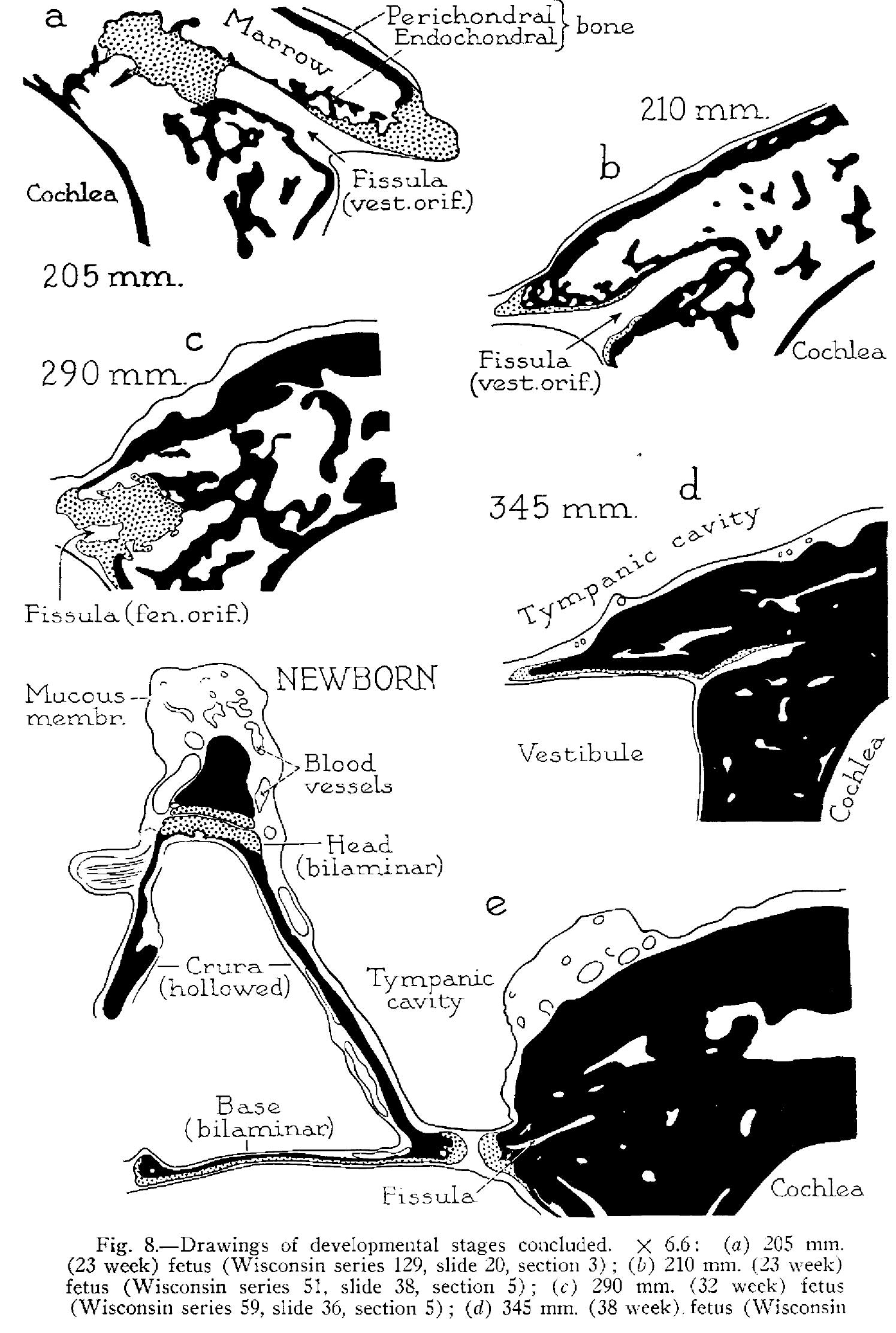 | 1,495 × 2,223 (335 KB) | Z8600021 (talk | contribs) |
You cannot overwrite this file.
File usage
The following 2 pages use this file: