File:Anson1948 fig02.jpg
Original file (1,280 × 1,232 pixels, file size: 241 KB, MIME type: image/jpeg)
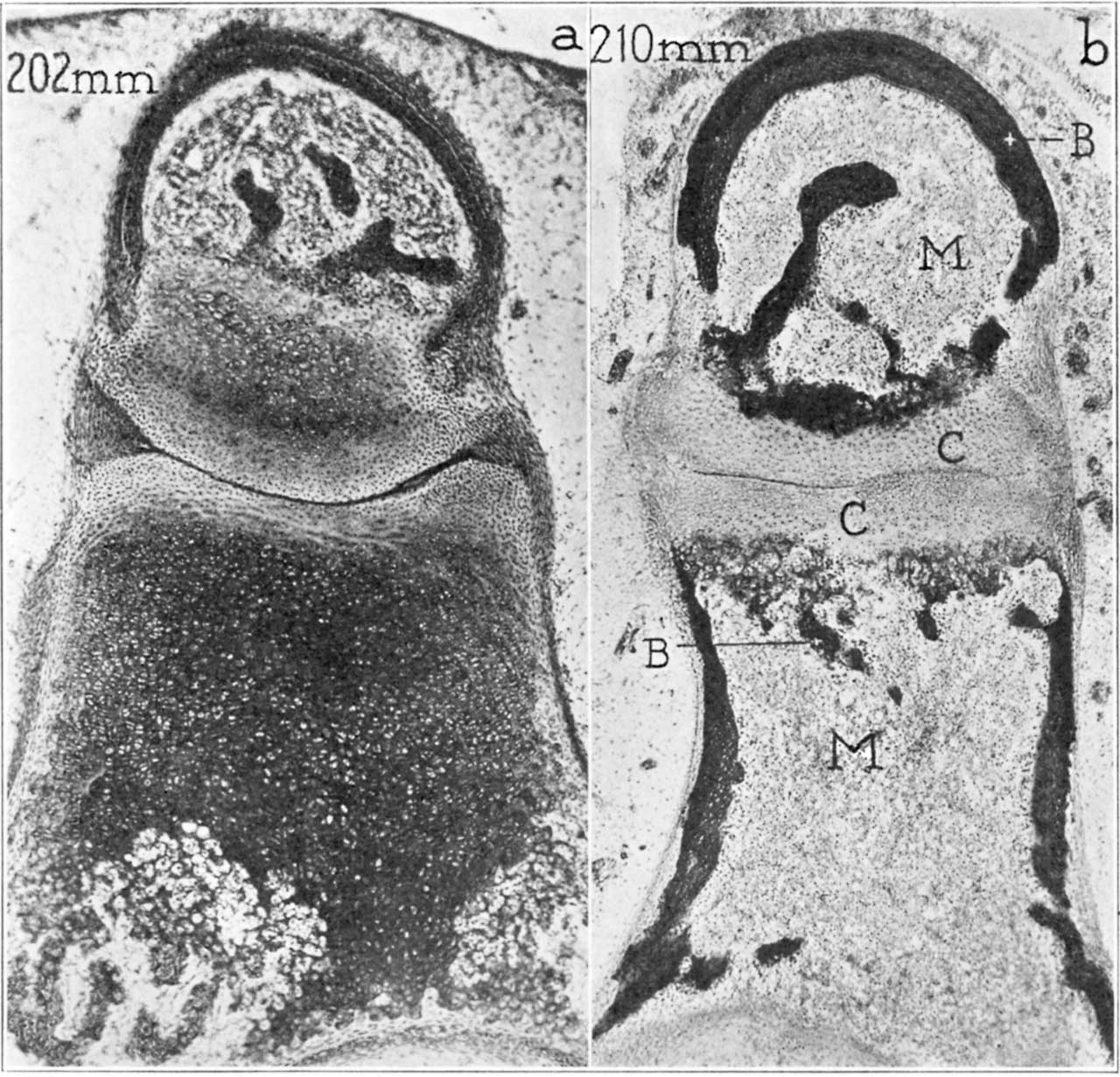
Fig. 2. Photomicrographs of 202 mm and 310 mm fetus stapes and incus
Photomicrographs of the neck and head of the stapes and of the lentieular process of the incus, showing erosion of the cartilage and its ultimate replacement (except where articular) by bone. x40 (a) 202 mm fetus (Wisconsin series 70 slide 37, section 6); (b) 310 mm fetus (Wisconsin series 51, slide 38, section 6).
Abbreviations: B indicates bone (perichondrial in the incus, endochondral in the head of the stapes; C, cartilage (of the articular plates of the ineus and stapes); M, marrow.
Reference
Anson BJ. and Cauldwell EW. Stapes, fissula ante fenestram and associated structures in man: V . From the fetus of 160 mm to term. (1948) 48(3): 263-300.
Cite this page: Hill, M.A. (2024, April 24) Embryology Anson1948 fig02.jpg. Retrieved from https://embryology.med.unsw.edu.au/embryology/index.php/File:Anson1948_fig02.jpg
- © Dr Mark Hill 2024, UNSW Embryology ISBN: 978 0 7334 2609 4 - UNSW CRICOS Provider Code No. 00098G
File history
Click on a date/time to view the file as it appeared at that time.
| Date/Time | Thumbnail | Dimensions | User | Comment | |
|---|---|---|---|---|---|
| current | 08:55, 15 October 2017 | | 1,280 × 1,232 (241 KB) | Z8600021 (talk | contribs) | |
| 08:48, 15 October 2017 |  | 1,576 × 1,811 (543 KB) | Z8600021 (talk | contribs) |
You cannot overwrite this file.
File usage
The following 2 pages use this file: