File:Alveolar-sac-01.jpg: Difference between revisions
(==Alveolar Sac== Source: modified from figure equation11.png also modified from (Hawgood & Clements 1990). http://herkules.oulu.fi/isbn9514270584/html/c273.html Category:Respiratory Category:Cartoon) |
mNo edit summary |
||
| (8 intermediate revisions by 2 users not shown) | |||
| Line 1: | Line 1: | ||
==Alveolar Sac== | ==Alveolar Sac== | ||
[[File:Respiratory_histology_03.jpg|thumb|300px|Alveous]] | |||
'''Alveolar Type I cell''' - (squamous alveolar) cells form the structure of an alveolar wall. | |||
'''Alveolar Type II cell''' - (great alveolar) cells secrete pulmonary {{surfactant}}. | |||
* {{surfactant}} is continuously released by exocytosis | |||
* lowers the surface tension of water and allows the membrane to separate | |||
* increases the capability to exchange gases. | |||
'''Macrophages''' - destroy foreign material and debris. | |||
Compare the above diagram structure with the [[:File:Respiratory_histology_03.jpg|alveoli histology]] and [[:File:Human lung inter-alveolar septum em01.jpg|electron microscope image]]. | |||
{{Respiratory Histology}} | |||
{{Respiratory Links}} | |||
===Reference=== | |||
Image Source: modified from figure equation11.png also modified from (Hawgood & Clements 1990). http://herkules.oulu.fi/isbn9514270584/html/c273.html | |||
{{Footer}} | |||
[[Category:Respiratory]] [[Category:Cartoon]] | [[Category:Respiratory]] [[Category:Cartoon]] | ||
Latest revision as of 22:03, 25 February 2019
Alveolar Sac

Alveolar Type I cell - (squamous alveolar) cells form the structure of an alveolar wall.
Alveolar Type II cell - (great alveolar) cells secrete pulmonary surfactant.
- surfactant is continuously released by exocytosis
- lowers the surface tension of water and allows the membrane to separate
- increases the capability to exchange gases.
Macrophages - destroy foreign material and debris.
Compare the above diagram structure with the alveoli histology and electron microscope image.
- Respiratory Histology: Bronchiole | Alveolar Duct | Alveoli | EM Alveoli septum | Alveoli Elastin | Trachea 1 | Trachea 2 | labeled lung | unlabeled lung | Respiratory Bronchiole | Lung Reticular Fibres | Nasal Inferior Concha | Nasal Respiratory Epithelium | Olfactory Region overview | Olfactory Region Epithelium | Histology Stains
Reference
Image Source: modified from figure equation11.png also modified from (Hawgood & Clements 1990). http://herkules.oulu.fi/isbn9514270584/html/c273.html
Cite this page: Hill, M.A. (2024, April 25) Embryology Alveolar-sac-01.jpg. Retrieved from https://embryology.med.unsw.edu.au/embryology/index.php/File:Alveolar-sac-01.jpg
- © Dr Mark Hill 2024, UNSW Embryology ISBN: 978 0 7334 2609 4 - UNSW CRICOS Provider Code No. 00098G
File history
Click on a date/time to view the file as it appeared at that time.
| Date/Time | Thumbnail | Dimensions | User | Comment | |
|---|---|---|---|---|---|
| current | 08:40, 25 August 2010 | 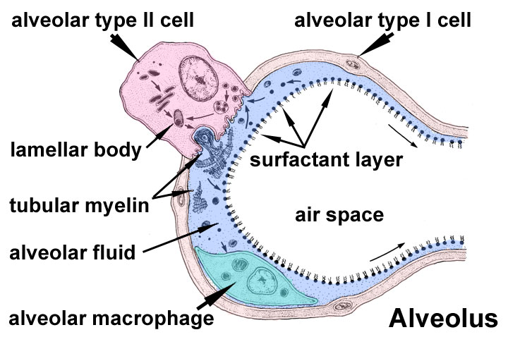 | 720 × 478 (73 KB) | S8600021 (talk | contribs) | ==Alveolar Sac== Source: modified from figure equation11.png also modified from (Hawgood & Clements 1990). http://herkules.oulu.fi/isbn9514270584/html/c273.html Category:Respiratory Category:Cartoon |
You cannot overwrite this file.
File usage
The following 15 pages use this file:
- 2010 Lecture 10
- 2011 Lab 5 - Fetal
- ANAT2241 Respiratory System
- ANAT2341 Lab 11 - Third Trimester
- ANAT2341 Lab 5 - Fetal
- ANAT2511 Respiratory System
- BGDA Practical 12 - Third Trimester
- Draft 2016
- Lecture - Respiratory Development
- Respiratory System - Postnatal
- Respiratory System Development
- SH Lecture - Respiratory System Development
- SH Practical - Respiratory
- Third Trimester
- File:Respiratory histology 03.jpg