File:Air blood barrier.gif
Air_blood_barrier.gif (397 × 517 pixels, file size: 178 KB, MIME type: image/gif)
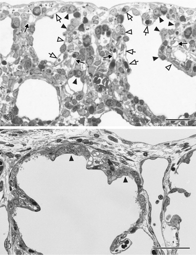
Air-blood barrier formation in Canalicular stage of Human Lung Development
The formation of the air-blood barrier in the human lung is a crucially important functional step in its development. This diagram displays the change in specialised epithelium which consequently forms this air-blood barrier.
Original Legend Text
Formation of the air–blood barrier. During the early canalicular stage (a, rat lung) the epithelium of the terminal airways is still cuboidal and glycogen-rich (closed arrowhead). Already a bit more proximal, the epithelium begins to flatten out (open arrowhead) and starts to form the first optimized future air–blood barriers. During the latter process, the capillaries of the mesenchyme (closed arrow) “move” towards the epithelium (open arrow). In humans (b), remnants of the cuboidal epithelium (closed arrowhead) are still present at the uttermost periphery of the gas exchange region at postnatal day 26, even if alveolarization had already started approximately 6 weeks earlier. This finding illustrates the large overlap between different phases of lung development, especially if peripheral and central parts are compared. Light microscopical images, bar 50 μm. (From Woods and Schittny 2016, by courtesy of Cambridge University Press, New York) [1]
Original source
Woods JC, Schittny JC (2016) Lung structure at preterm and term birth. In: Jobe AH, Whitsett JA, Abman SH (eds) Fetal lung development - clinical correlates & future technologies. Cambridge University Press, New York, pp 126–140
Copyright
This article is distributed under the terms of the Creative Commons Attribution 4.0 International License (http://creativecommons.org/licenses/by/4.0/), which permits unrestricted use, distribution, and reproduction in any medium, provided you give appropriate credit to the original author(s) and the source, provide a link to the Creative Commons license, and indicate if changes were made.
Reference
- ↑ Schittny, J. C. (2017). Development of the lung. Cell and Tissue Research, 367(3), 427–444. http://doi.org/10.1007/s00441-016-2545-0
File history
Click on a date/time to view the file as it appeared at that time.
| Date/Time | Thumbnail | Dimensions | User | Comment | |
|---|---|---|---|---|---|
| current | 10:13, 5 October 2017 | | 397 × 517 (178 KB) | Z5059373 (talk | contribs) |
You cannot overwrite this file.
File usage
The following 2 pages use this file: