File:Adult urinary bladder.jpg
From Embryology
Size of this preview: 698 × 600 pixels. Other resolution: 1,164 × 1,000 pixels.
Original file (1,164 × 1,000 pixels, file size: 148 KB, MIME type: image/jpeg)
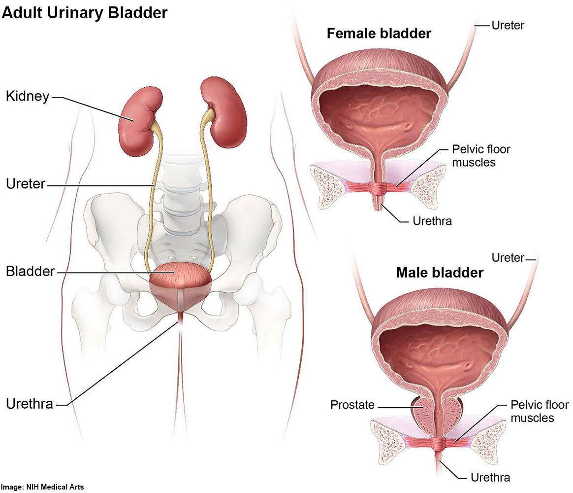
Adult Urinary Bladder
An illustration of the male and female human urinary system, including kidneys, bladder, ureter and urethra, pelvic floor muscles and the prostate.
Reference
Credit: NIH Medical Arts
Cite this page: Hill, M.A. (2024, April 25) Embryology Adult urinary bladder.jpg. Retrieved from https://embryology.med.unsw.edu.au/embryology/index.php/File:Adult_urinary_bladder.jpg
- © Dr Mark Hill 2024, UNSW Embryology ISBN: 978 0 7334 2609 4 - UNSW CRICOS Provider Code No. 00098G
File history
Click on a date/time to view the file as it appeared at that time.
| Date/Time | Thumbnail | Dimensions | User | Comment | |
|---|---|---|---|---|---|
| current | 14:52, 9 March 2017 | | 1,164 × 1,000 (148 KB) | Z8600021 (talk | contribs) | ==Adult Urinary Bladder== |
You cannot overwrite this file.
File usage
The following page uses this file: