File:Adult heart outflow tract CT02.jpg
Original file (747 × 747 pixels, file size: 65 KB, MIME type: image/jpeg)
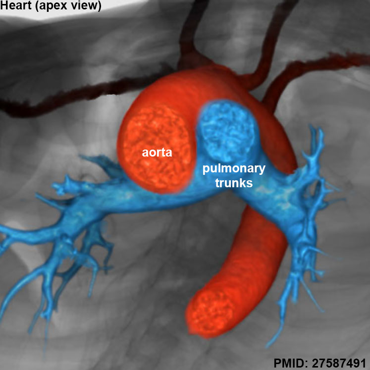
Adult Heart Outflow Tract
Reconstruction seen in short-axis projection from the cardiac apex. Shows the spiraling nature of the intrapericardial arterial trunks.
Red - aorta
Blue - pulmonary trunk
See also unlabeled image version
Reference
<pubmed>27587491</pubmed>
https://www.ncbi.nlm.nih.gov/pmc/articles/PMC5011314/
http://journals.sagepub.com/doi/abs/10.1177/2150135116651114
PMID 27587491
Copyright
© The Author(s) 2016
https://creativecommons.org/licenses/by/3.0/
Figure 12. cropped and resized, labeled with PMID. Text above modified from figure legend.
Cite this page: Hill, M.A. (2024, April 25) Embryology Adult heart outflow tract CT02.jpg. Retrieved from https://embryology.med.unsw.edu.au/embryology/index.php/File:Adult_heart_outflow_tract_CT02.jpg
- © Dr Mark Hill 2024, UNSW Embryology ISBN: 978 0 7334 2609 4 - UNSW CRICOS Provider Code No. 00098G
File history
Click on a date/time to view the file as it appeared at that time.
| Date/Time | Thumbnail | Dimensions | User | Comment | |
|---|---|---|---|---|---|
| current | 15:03, 29 January 2017 | | 747 × 747 (65 KB) | Z8600021 (talk | contribs) | ==Adult Heart Outflow Tract== Reconstruction seen in short-axis projection from the cardiac apex. Shows the spiraling nature of the intrapericardial arterial trunks. Red - aorta Blue - pulmonary trunk See also [[:File:Adult heart outflow tract CT... |
You cannot overwrite this file.
File usage
There are no pages that use this file.