Fetal Development - 18 Weeks: Difference between revisions
mNo edit summary |
mNo edit summary |
||
| Line 11: | Line 11: | ||
|-bgcolor="F5FAFF" | |-bgcolor="F5FAFF" | ||
| | | | ||
* '''Prediction of newborn birth weight based on the estimation at 20-24 weeks of gestation'''<ref><pubmed>21056312</pubmed></ref> "We propose an accurate, simple, and easy formula to better assess the newborn birth weight at mid-pregnancy for the Asian population. Mid-pregnancy BMI was a more significant factor for birth weight estimation than other maternal weight factors in this study." | * '''Prediction of newborn birth weight based on the estimation at 20-24 weeks of gestation'''<ref name=PMID21056312><pubmed>21056312</pubmed></ref> "We propose an accurate, simple, and easy formula to better assess the newborn birth weight at mid-pregnancy for the Asian population. Mid-pregnancy BMI was a more significant factor for birth weight estimation than other maternal weight factors in this study." | ||
|} | |} | ||
==Neural== | |||
===Spinal Cord=== | |||
An ultrasound study of the position of the spinal cord conus medullaris at 18-22 weeks (20 to 24 weeks ((GA}}) showed that it ended adjacent to vertebrae L2, L2-3 vertebral space, and L3 (73/78, 93%).<ref name=PMID20582935><pubmed>20582935</pubmed></ref> The conus medullaris (Latin, "medullary cone") is the tapered, lower end of the spinal cord. | |||
:'''Links:''' [[Neural - Spinal Cord Development|Spinal Cord Development]] | |||
==Thymus Histology== | ==Thymus Histology== | ||
Latest revision as of 08:27, 23 March 2016
| Embryology - 19 Apr 2024 |
|---|
| Google Translate - select your language from the list shown below (this will open a new external page) |
|
العربية | català | 中文 | 中國傳統的 | français | Deutsche | עִברִית | हिंदी | bahasa Indonesia | italiano | 日本語 | 한국어 | မြန်မာ | Pilipino | Polskie | português | ਪੰਜਾਬੀ ਦੇ | Română | русский | Español | Swahili | Svensk | ไทย | Türkçe | اردو | ייִדיש | Tiếng Việt These external translations are automated and may not be accurate. (More? About Translations) |
Introduction
Images on this current page show thymus development histology occurring during the 18 week post-fertilization age or 20 week gestational age (GA) second trimester stage of development.
| Fetal Links: fetal | Week 10 | Week 12 | second trimester | third trimester | fetal neural | Fetal Blood Sampling | fetal growth restriction | birth | birth weight | preterm birth | Developmental Origins of Health and Disease | macrosomia | BGD Practical | Medicine Lecture | Science Lecture | Lecture Movie | Category:Human Fetus | Category:Fetal | |||
|
Some Recent Findings
|
Neural
Spinal Cord
An ultrasound study of the position of the spinal cord conus medullaris at 18-22 weeks (20 to 24 weeks ((GA}}) showed that it ended adjacent to vertebrae L2, L2-3 vertebral space, and L3 (73/78, 93%).[2] The conus medullaris (Latin, "medullary cone") is the tapered, lower end of the spinal cord.
- Links: Spinal Cord Development
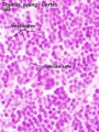
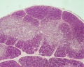
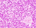
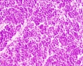
Thymus Histology
- Thymus Histology: Fetal Thymus overview | Fetal Thymus Medulla | Fetal Thymus Cortex | Adult Thymus | unlabeled fetal overview | unlabeled fetal medulla |unlabeled fetal thymic corpuscle |unlabeled fetal cortex | unlabeled adult overview | Category:Thymus | Immune System Development
The developing fetal thymus shown below is from a 20 week gestational age (GA), 18 week post-fertilization age, or second trimester stage of development.
Note, there is no specific event occurring in thymus growth during week 18, this happens to be the period available to show fatal histology.
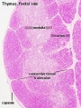
Fetal Thymus overview
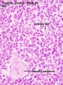
Fetal Thymus Medulla
Fetal Thymus Cortex
unlabeled fetal overview
unlabeled fetal medulla

unlabeled fetal thymic corpuscle
unlabeled fetal cortex


References
Reviews
<pubmed></pubmed>
Articles
<pubmed>21623033</pubmed>
Search PubMed
Note: Week 18 post-fertilization age (used throughout this current website) is Week 20 gestational age (LMP). Searches for clinical Week 20 gestational age will match this post-fertilization age.
Search PubMed Now: week 20 fetus | second trimester fetus
Glossary Links
- Glossary: A | B | C | D | E | F | G | H | I | J | K | L | M | N | O | P | Q | R | S | T | U | V | W | X | Y | Z | Numbers | Symbols | Term Link
Cite this page: Hill, M.A. (2024, April 19) Embryology Fetal Development - 18 Weeks. Retrieved from https://embryology.med.unsw.edu.au/embryology/index.php/Fetal_Development_-_18_Weeks
- © Dr Mark Hill 2024, UNSW Embryology ISBN: 978 0 7334 2609 4 - UNSW CRICOS Provider Code No. 00098G