Fetal Development
| Embryology - 19 Apr 2024 |
|---|
| Google Translate - select your language from the list shown below (this will open a new external page) |
|
العربية | català | 中文 | 中國傳統的 | français | Deutsche | עִברִית | हिंदी | bahasa Indonesia | italiano | 日本語 | 한국어 | မြန်မာ | Pilipino | Polskie | português | ਪੰਜਾਬੀ ਦੇ | Română | русский | Español | Swahili | Svensk | ไทย | Türkçe | اردو | ייִדיש | Tiếng Việt These external translations are automated and may not be accurate. (More? About Translations) |
Introduction

| <mediaplayer width='285' height='320' image="http://php.med.unsw.edu.au/embryology/images/f/ff/Fetal_growth_icon.jpg">File:fetal growth.mp4</mediaplayer> | This page shows some key events of human development during the fetal period (weeks 9 to 37) following fertilization. The long Fetal period (4x the embryonic period) is a time of extensive growth in size and mass as well as ongoing differentiation of organ systems established in the embryonic period. Clinically this period is generally described as the Second Trimester and Third Trimester. Many of the critical measurements of growth are now carried out by ultrasound and this period ends at birth.
Many different systems formed in the embryonic period (organogenesis) grow and differentiate further during the fetal period and do so at different times. For example, the brain continues to grow and develop extensively during this period (and postnatally), the respiratory system differentiates (and completes only just before birth), the urogenital system further differentiates between male/female, endocrine and gastrointestinal tract begins to function. Also consider the systems (respiratory, cardiac, neural) that will still not have their final organization and function determined until after birth. |
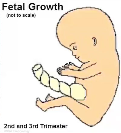
| Changing fetal proportions, not size growth. | Use the links below to get more detailed information about this period of development. |
| Fetal Links: fetal | Week 10 | Week 12 | second trimester | third trimester | fetal neural | Fetal Blood Sampling | fetal growth restriction | birth | birth weight | preterm birth | Developmental Origins of Health and Disease | macrosomia | BGD Practical | Medicine Lecture | Science Lecture | Lecture Movie | Category:Human Fetus | Category:Fetal | |||
|
- Fetal Graphs: Crown-Rump Length (CRL) | Third trimester CRL | Head Circumference | Head Circumference 2nd Trimester | Liver Weight | Pancreas Weight | Thymus Weight | Small Intestine Length | Large Intestine Length | Length and Weight Changes | Fetal Development
Some Recent Findings

|
| More recent papers |
|---|
 This table allows an automated computer search of the external PubMed database using the listed "Search term" text link.
More? References | Discussion Page | Journal Searches | 2019 References | 2020 References Search term: Fetal Development <pubmed limit=5>Fetal+Development</pubmed> |
Reading
|
|

Second Trimester

- Second Trimester
- Week 12 - CRL 85 mm, femur length 15 mm, biparietal diameter 25 mm.
Begin by working through the features present in the early 10 week female fetus. Then look in detail at the head development in a 12 week fetus.


|

|

|

|
Then look in detail at the head development in a 12 week fetus showing both forms of ossification in the skull.

|

|

|
Fetal Head Growth
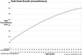
Second and third trimesters
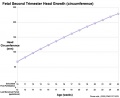
Second trimester


Third Trimester


- Vibration acoustically of maternal abdominal wall induces startle respone in fetus.
- Month 7 - respiratory bronchioles proliferate and end in alveolar ducts and sacs.
- Week 37 to 38 Birth.
- Links: Third Trimester
Fetal Endocrine

Pituitary Hormones
- HPA axis established by week 20
- Pituitary functional throughout fetal development
Thyroid Hormone
- required for metabolic activity, also in the newborn
- important for neural development
Parathyroid Hormone
- newborn has total calcium levels (approx 20 grams) accumulated mainly in the 3rd trimester (weeks 28–40)
- fetal parathyroid hormone (PTH) potentially available from 10–12 weeks and PTH does not cross the placenta
- fetus relatively hypercalcemic, active transplacental transport of Ca2+ to fetus
- maternal serum - calcium ions (Ca2+), inorganic phosphate (Pi) and PTH concentrations are within the non-pregnant normal range throughout pregnancy.
- maternal bone turnover increases in the 3rd trimester.
(Based on Endocrinology - Materno—fetal calcium balance)
Pancreatic Hormones
- maternal diabetes can affect fetal pancreas development (increase in fetal islet beta cells).
Gonadal Hormones
- testosterone - required during fetal development for external genital development and internal genital tract in male.
- estrogens - secreted inactive precursor converted to active form by placenta.
- Links: Endocrine System Development | Endocrinology - Control of steroid production in the fetal gonads | Neuroscience - The Effect of Sex Hormones on Neural Circuitry
Gastrointestinal Tract

|

|
Fetal developmental features include: the growth and rotation of intestines initially herniated outside the ventral body wall; changes in mesenteries; development of the blood supply and tract wall.
The initial functions of the tract with amionic fluid swallowing and the accumulation of both secretions and swallowed components within the large intestine as meconium.
Fetal Surgical Procedures
There are a range of fatal abnormalities that are potentially amenable to surgical intervention, see recent review link Maternal-Fetal Surgical Procedures.
Some examples include:
- Cardiac Malformations
- Congenital Diaphragmatic Hernia
- Myelomeningocele/Spina Bifida
- Obstructive Uropathy
- Sacrococcygeal Teratoma
- Thoracic Lesions
- Twin-Twin Transfusion Syndrome
- Links: Maternal-Fetal Surgical Procedures. Walsh WF, Chescheir NC, Gillam-Krakauer M, et al. Rockville (MD): Agency for Healthcare Research and Quality (US); 2011 Apr. (Comparative Effectiveness Technical Briefs, No. 5.) Report | Comparative Effectiveness Research, Health Care
References
Journals
Reviews
- Fetal assessment during pregnancy. Farley D, Dudley DJ. Pediatr Clin North Am. 2009 Jun;56(3):489-504, Table of Contents. Review. PMID: 19501688
- Regimens of fetal surveillance for impaired fetal growth. Grivell RM, Wong L, Bhatia V. Cochrane Database Syst Rev. 2009 Jan 21;(1):CD007113. Review. PMID: 19160321
Articles
Search PubMed
Search Pubmed: human fetal development | fetal development | Second Trimester | Third Trimester
Glossary Links
- Glossary: A | B | C | D | E | F | G | H | I | J | K | L | M | N | O | P | Q | R | S | T | U | V | W | X | Y | Z | Numbers | Symbols | Term Link
Cite this page: Hill, M.A. (2024, April 19) Embryology Fetal Development. Retrieved from https://embryology.med.unsw.edu.au/embryology/index.php/Fetal_Development
- © Dr Mark Hill 2024, UNSW Embryology ISBN: 978 0 7334 2609 4 - UNSW CRICOS Provider Code No. 00098G