Episcopic Fluorescence Image Capture: Difference between revisions
mNo edit summary |
mNo edit summary |
||
| Line 13: | Line 13: | ||
|-bgcolor="F5FAFF" | |-bgcolor="F5FAFF" | ||
| | | | ||
* '''Spatial | * '''Spatial change of cruciate ligaments in rat embryo knee joint by three-dimensional reconstruction'''<ref name=PMID26098761><pubmed>26098761</pubmed></ref> "This study aimed to analyze the spatial developmental changes of rat cruciate ligaments by three-dimensional (3D) reconstruction using episcopic fluorescence image capture (EFIC). Cruciate ligaments of Wister rat embryos between embryonic day (E) 16 and E20 were analyzed. Samples were sectioned and visualized using EFIC. 3D reconstructions were generated using Amira software. The length of the cruciate ligaments, distances between attachment points to femur and tibia, angles of the cruciate ligaments and the cross angle of the cruciate ligaments were measured. The shape of cruciate ligaments was clearly visible at E17. The lengths of the anterior cruciate ligament (ACL) and posterior cruciate ligament (PCL) increased gradually from E17 to E19 and drastically at E20. Distances between attachment points to the femur and tibia gradually increased. The ACL angle and PCL angle gradually decreased. The cross angle of the cruciate ligaments changed in three planes. The primordium of the 3D structure of rat cruciate ligaments was constructed from the early stage, with the completion of the development of the structures occurring just before birth." | ||
* '''A detailed comparison of mouse and human cardiac development'''<ref name=PMID25167202><pubmed>25167202</pubmed></ref> "Mouse mutants are used to model human congenital cardiovascular disease. Few studies exist comparing normal cardiovascular development in mice vs. humans. We carried out a systematic comparative analysis of mouse and human fetal cardiovascular development. Episcopic fluorescence image capture (EFIC) was performed on 66 wild-type mouse embryos from embryonic day (E) 9.5 to birth; 2-dimensional and 3-dimensional datasets were compared with EFIC and magnetic resonance images from a study of 52 human fetuses (Carnegie stage 13-23). Time course of atrial, ventricular, and outflow septation were outlined and followed a similar sequence in both species. Bilateral venae cavae and prominent atrial appendages were seen in the mouse fetus; in human fetuses, atrial appendages were small, and a single right superior vena cava was present. In contrast to humans with separate pulmonary vein orifices, a pulmonary venous confluence with one orifice enters the left atrium in mice." | |||
|} | |} | ||
{| class="wikitable mw-collapsible mw-collapsed" | {| class="wikitable mw-collapsible mw-collapsed" | ||
Revision as of 12:12, 17 August 2016
| Embryology - 23 Apr 2024 |
|---|
| Google Translate - select your language from the list shown below (this will open a new external page) |
|
العربية | català | 中文 | 中國傳統的 | français | Deutsche | עִברִית | हिंदी | bahasa Indonesia | italiano | 日本語 | 한국어 | မြန်မာ | Pilipino | Polskie | português | ਪੰਜਾਬੀ ਦੇ | Română | русский | Español | Swahili | Svensk | ไทย | Türkçe | اردو | ייִדיש | Tiếng Việt These external translations are automated and may not be accurate. (More? About Translations) |
Introduction
Episcopic Fluorescence Image Capture (EFIC). A microscopic imaging technique that serially sections embedded biological specimens and photographs the tissue autofluorescence (epifluorescence) from the block surface. This generates an in register 2D image stack. The technique was described for the study of transgenic mice in 2002.[1]
Some Recent Findings
|
| More recent papers |
|---|
 This table allows an automated computer search of the external PubMed database using the listed "Search term" text link.
More? References | Discussion Page | Journal Searches | 2019 References | 2020 References Search term: Episcopic Fluorescence Image Capture <pubmed limit=5>Episcopic Fluorescence Image Capture</pubmed> |
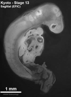
Stage 13
<html5media height="410" width="290">File:Stage 13 EFIC01.mp4</html5media>
Click Here to play on mobile device
See also full size and labelled version on the Stage 13 EFIC movie page.
References
Reviews
<pubmed></pubmed> <pubmed></pubmed> <pubmed></pubmed> <pubmed></pubmed>
Articles
<pubmed>20503356</pubmed> <pubmed></pubmed> <pubmed></pubmed> <pubmed></pubmed>
Search PubMed
Search Pubmed: Episcopic Fluorescence Image Capture
Terms
External Links
External Links Notice - The dynamic nature of the internet may mean that some of these listed links may no longer function. If the link no longer works search the web with the link text or name. Links to any external commercial sites are provided for information purposes only and should never be considered an endorsement. UNSW Embryology is provided as an educational resource with no clinical information or commercial affiliation.
Glossary Links
- Glossary: A | B | C | D | E | F | G | H | I | J | K | L | M | N | O | P | Q | R | S | T | U | V | W | X | Y | Z | Numbers | Symbols | Term Link
Cite this page: Hill, M.A. (2024, April 23) Embryology Episcopic Fluorescence Image Capture. Retrieved from https://embryology.med.unsw.edu.au/embryology/index.php/Episcopic_Fluorescence_Image_Capture
- © Dr Mark Hill 2024, UNSW Embryology ISBN: 978 0 7334 2609 4 - UNSW CRICOS Provider Code No. 00098G