Endoderm Development Movie: Difference between revisions
mNo edit summary |
mNo edit summary |
||
| Line 17: | Line 17: | ||
|} | |} | ||
'''Links:''' [[Media:Endoderm 003.mp4|MP4 labeled version]] | [[Media:Endoderm 002.mp4|MP4 unlabeled version]] | [[Media:Endoderm 001.mp4|MP4 large unlabeled version]] | [[Gastrointestinal Tract Development]] | [[ | '''Links:''' [[Media:Endoderm 003.mp4|MP4 labeled version]] | [[Media:Endoderm 002.mp4|MP4 unlabeled version]] | [[Media:Endoderm 001.mp4|MP4 large unlabeled version]] | [[Gastrointestinal Tract Development]] | [[Lecture_-_Gastrointestinal_Development|Lecture - Endoderm Development]] | [[Movies#Gastrointestinal_Tract|Gastrointestinal Tract Movies]] | ||
{| | {| | ||
Revision as of 09:26, 26 April 2013
| Embryology - 18 Apr 2024 |
|---|
| Google Translate - select your language from the list shown below (this will open a new external page) |
|
العربية | català | 中文 | 中國傳統的 | français | Deutsche | עִברִית | हिंदी | bahasa Indonesia | italiano | 日本語 | 한국어 | မြန်မာ | Pilipino | Polskie | português | ਪੰਜਾਬੀ ਦੇ | Română | русский | Español | Swahili | Svensk | ไทย | Türkçe | اردو | ייִדיש | Tiếng Việt These external translations are automated and may not be accurate. (More? About Translations) |
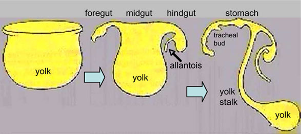
| <mediaplayer width='300' height='320' image="http://embryology.med.unsw.edu.au/embryology/images/a/af/Endoderm_002_icon.jpg">File:Endoderm 003.mp4</mediaplayer> | This animation shows the early development of endoderm forming the gastrointestinal tract, yolk sac and allantois. The movie starts approximately week 3 and continues through week 4.
Yellow shows the general lining of the yolk sac (bottom), continuous with the endoderm of the trilaminar embryonic disc (top) during week 3. As the trilaminar disc folds in this week, the foregut and hindgut regions become separated from the external yolk sac. The midgut region remains open to the yolk sac and will separate later. Foregut - Begins at the buccopharyngeal membrane, the foregut region in the head is now called the pharynx. At the lower end of the pharynx a ventral bud forms, that will later form the respiratory tract. Beneath this region the tube grows rapidly forming a dilation of the tube, that will later form the stomach. Beneath this region is the boundary of the foregut and ventrally lies the transverse septum. Midgut - Broadly open to the external yolk sac then with continued folding narrows to a "tube-like" connection the yolk stalk. This stalk will later degenerate and all connection will normally be lost. The yolk sac is pushed to the periphery by the growing amniotic sac, with its connecting yolk stalk in the umbilicus region. The midgut region also grows in length forming a loop lying outside the ventral body wall. Hindgut - The loop of midgut renters the body and the ventral portion of the hindgut extends as a blind-ended tube, or diverticulum, into the connecting stalk. This endoderm extension can be seen in histological sections of the initial placental cord and is called the allantois. The hindgut extends caudal (tailward) ending at the cloacal membrane. |
Links: MP4 labeled version | MP4 unlabeled version | MP4 large unlabeled version | Gastrointestinal Tract Development | Lecture - Endoderm Development | Gastrointestinal Tract Movies

|
|
Glossary Links: A | B | C | D | E | F | G | H | I | J | K | L | M | N | O | P | Q | R | S | T | U | V | W | X | Y | Z | Numbers | Symbols | Movies
Cite this page: Hill, M.A. (2024, April 18) Embryology Endoderm Development Movie. Retrieved from https://embryology.med.unsw.edu.au/embryology/index.php/Endoderm_Development_Movie
- © Dr Mark Hill 2024, UNSW Embryology ISBN: 978 0 7334 2609 4 - UNSW CRICOS Provider Code No. 00098G