Endocrine - Thymus Development: Difference between revisions
mNo edit summary |
|||
| Line 17: | Line 17: | ||
==Some Recent Findings== | ==Some Recent Findings== | ||
[[File:Human embryo thymus and parathyroid 01.jpg|thumb|Human Embryo (week 6 - 8) | [[File:Human embryo thymus and parathyroid 01.jpg|thumb|Human Embryo (week 6 - 8){{#pmid:21203493|PMID21203493}}]] | ||
{| | {| | ||
|-bgcolor="F5FAFF" | |-bgcolor="F5FAFF" | ||
| Line 66: | Line 66: | ||
| [[:File:Stage_22_image_071.jpg|D1]] Developing Human Thymus ([[Carnegie stage 22|stage 22]]) | | [[:File:Stage_22_image_071.jpg|D1]] Developing Human Thymus ([[Carnegie stage 22|stage 22]]) | ||
|} | |} | ||
==Thymus Vasculature== | |||
Like all endocrine organs the thymus is eventually richly vascularised, development has been previously summarised.{{#pmid:7445948|PMID7445948}} | |||
* {{GA}} week 10 - initial blood supply. | |||
* {{GA}} week 12 - interlobular septa blood spaces late normoblasts and granulocytes increase, cortical and medullary vasculature increases. | |||
* {{GA}} week 16 - nerve bundles accompany arteries and veins. | |||
* {{GA}} week 20 to 24 - radial cortical capillaries drain into capsular venules. The arterioles give rise to a series of radial cortical capillaries and less regular vessels to the medulla. | |||
* {{GA}} week 28 to 40 - vascular thymic supply markedly increases and cortical capillaries can anastomose. | |||
==Thymus Involution== | ==Thymus Involution== | ||
| Line 77: | Line 86: | ||
===Reviews=== | ===Reviews=== | ||
{{#pmid:19582736}} | |||
{{#pmid:18304000}} | |||
{{#pmid:17876091}} | |||
{{#pmid:16448532}} | |||
===Articles=== | ===Articles=== | ||
{{#pmid:17625108}} | |||
{{#pmid:15057786}} | |||
{{#pmid:11742403}} | |||
===Search PubMed=== | ===Search PubMed=== | ||
Revision as of 15:10, 23 February 2018
| Embryology - 24 Apr 2024 |
|---|
| Google Translate - select your language from the list shown below (this will open a new external page) |
|
العربية | català | 中文 | 中國傳統的 | français | Deutsche | עִברִית | हिंदी | bahasa Indonesia | italiano | 日本語 | 한국어 | မြန်မာ | Pilipino | Polskie | português | ਪੰਜਾਬੀ ਦੇ | Română | русский | Español | Swahili | Svensk | ไทย | Türkçe | اردو | ייִדיש | Tiếng Việt These external translations are automated and may not be accurate. (More? About Translations) |
Introduction

The thymus has two origins for the lymphoid thymocytes and the thymic epithelial cells. The thymic epithelium begins as two flask-shape endodermal diverticula that form from the third pharyngeal pouch and extend lateralward and backward into the surrounding mesoderm and neural crest-derived mesenchyme in front of the ventral aorta. The immune system T cells are essential for responses against infections and much research concerns the postnatal development of T cells within the thymus.
Stieda in 1881[1] was the first to observe that the thymus gland originated from a visceral (pharyngeal) pouch (endoderm).
This current page relates to the endocrine role of the thymus, for more detailed description of this organ development see Thymus Development.
| Immune Links: immune | blood | spleen | thymus | lymphatic | lymph node | Antibody | Med Lecture - Lymphatic Structure | Med Practical | Immune Movies | vaccination | bacterial infection | Abnormalities | Category:Immune | ||
|
Some Recent Findings

|
| More recent papers |
|---|
 This table allows an automated computer search of the external PubMed database using the listed "Search term" text link.
More? References | Discussion Page | Journal Searches | 2019 References | 2020 References Search term: Thymus Embryology <pubmed limit=5>Thymus Embryology</pubmed> |
Thymus Hormones
Thymus produces self-hormones
- thymulin
- thymosin
- thymopentin
- thymus humoral factor
Thymus Development


- Endoderm - third pharyngeal pouch
- Week 6 - diverticulum elongates, hollow then solid, ventral cell proliferation
- Thymic primordia - surrounded by neural crest mesenchyme, epithelia/mesenchyme interaction
- Thymus - bone-marrow lymphocyte precursors become thymocytes, and subsequently mature into T lymphocytes (T cells)
- Thymus hormones - thymosins stimulate the development and differentiation of T lymphocytes

|

|
| B2 Pharyngeal Arch Pouches 3 and 4 (stage 13) | D1 Developing Human Thymus (stage 22) |
Thymus Vasculature
Like all endocrine organs the thymus is eventually richly vascularised, development has been previously summarised.[6]
- GA week 10 - initial blood supply.
- GA week 12 - interlobular septa blood spaces late normoblasts and granulocytes increase, cortical and medullary vasculature increases.
- GA week 16 - nerve bundles accompany arteries and veins.
- GA week 20 to 24 - radial cortical capillaries drain into capsular venules. The arterioles give rise to a series of radial cortical capillaries and less regular vessels to the medulla.
- GA week 28 to 40 - vascular thymic supply markedly increases and cortical capillaries can anastomose.
Thymus Involution
A postnatal process defined as a decrease in the size, weight and activity of the gland with advancing age. In a recent review[7], thymic involution was described as a result of high levels of circulating sex hormones, in particular during puberty, and a lower population of precursor cells from the bone marrow and finally changes in the thymic microenvironment.
References
- ↑ Stieda L (1881) Untersuchungen über die Entwickelung der Glandular Thymus, Glandular Thyreoidea, und Glandular carotidica. Leipzig, Engelmann p38.
- ↑ 2.0 2.1 Liu Z, Farley A, Chen L, Kirby BJ, Kovacs CS, Blackburn CC & Manley NR. (2010). Thymus-associated parathyroid hormone has two cellular origins with distinct endocrine and immunological functions. PLoS Genet. , 6, e1001251. PMID: 21203493 DOI. Cite error: Invalid
<ref>tag; name 'PMID21203493' defined multiple times with different content - ↑ <pubmed>28279759</pubmed>
- ↑ <pubmed>23571219</pubmed>
- ↑ <pubmed>20644572</pubmed>
- ↑ Ghali WM, Abdel-Rahman S, Nagib M & Mahran ZY. (1980). Intrinsic innervation and vasculature of pre- and post-natal human thymus. Acta Anat (Basel) , 108, 115-23. PMID: 7445948
- ↑ <pubmed>20354268 </pubmed>
Reviews
Anderson G, Jenkinson EJ & Rodewald HR. (2009). A roadmap for thymic epithelial cell development. Eur. J. Immunol. , 39, 1694-9. PMID: 19582736 DOI.
Rodewald HR. (2008). Thymus organogenesis. Annu. Rev. Immunol. , 26, 355-88. PMID: 18304000 DOI.
Nowell CS, Farley AM & Blackburn CC. (2007). Thymus organogenesis and development of the thymic stroma. Methods Mol. Biol. , 380, 125-62. PMID: 17876091 DOI.
Holländer G, Gill J, Zuklys S, Iwanami N, Liu C & Takahama Y. (2006). Cellular and molecular events during early thymus development. Immunol. Rev. , 209, 28-46. PMID: 16448532 DOI.
Articles
Itoi M, Tsukamoto N, Yoshida H & Amagai T. (2007). Mesenchymal cells are required for functional development of thymic epithelial cells. Int. Immunol. , 19, 953-64. PMID: 17625108 DOI.
Blackburn CC & Manley NR. (2004). Developing a new paradigm for thymus organogenesis. Nat. Rev. Immunol. , 4, 278-89. PMID: 15057786 DOI.
Rodewald HR, Paul S, Haller C, Bluethmann H & Blum C. (2001). Thymus medulla consisting of epithelial islets each derived from a single progenitor. Nature , 414, 763-8. PMID: 11742403 DOI.
Search PubMed
Search Pubmed: thymus development
Additional Images
Historic Images
| Historic Disclaimer - information about historic embryology pages |
|---|
|
Sudler, MT. The Development of the Nose and of the Pharynx and its Derivatives in Man. (1902) Amer. J. Anat 1:391–416. Thymus Gland
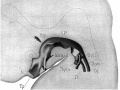
Fig. 8. Model of pharynx embryo CXLIV
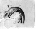
Fig. 9. Model of pharynx embryo XLIII
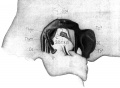
Fig. 10. Lateral view of embryo XXII


Adult Histology
Terms
- Hassall's corpuscle - thymic corpuscle.
- Thymic corpuscle (=Hassall's corpuscle) a mass of concentric epithelioreticular cells found in the thymus. The number present and size tend to increase with thymus age. (see classical description of Hammar, J. A. 1903 Zur Histogenese und Involution der Thymusdriise. Anat. Anz., 27: 1909 Fiinfzig Jahre Thymusforschung. Ergebn. Anat. Entwickl-gesch. 19: 1-274.)
- thymic epitheliocytes - reticular cells located in the thymus cortex that ensheathe the cortical capillaries, creating and maintain the microenvironment necessary for the development of T-lymphocytes in the cortex.
- T lymphocyte (cell) - named after thymus, where they develop, the active cell is responsible for cell-mediated immunity. (More? Electron micrographs of nonactivate and activated lymphocytes)
Glossary Links
- Glossary: A | B | C | D | E | F | G | H | I | J | K | L | M | N | O | P | Q | R | S | T | U | V | W | X | Y | Z | Numbers | Symbols | Term Link
Cite this page: Hill, M.A. (2024, April 24) Embryology Endocrine - Thymus Development. Retrieved from https://embryology.med.unsw.edu.au/embryology/index.php/Endocrine_-_Thymus_Development
- © Dr Mark Hill 2024, UNSW Embryology ISBN: 978 0 7334 2609 4 - UNSW CRICOS Provider Code No. 00098G