Endocrine - Pineal Development: Difference between revisions
mNo edit summary |
mNo edit summary |
||
| Line 177: | Line 177: | ||
{{Historic Disclaimer}} | {{Historic Disclaimer}} | ||
<gallery> | <gallery> | ||
File:Keith1902 fig017.jpg
|Keith1902 |
File:Keith1902 fig017.jpg
|Keith1902 Fig 17 | ||
File:Keith1902 fig167.jpg
|Keith1902 | File:Keith1902 fig167.jpg
|Keith1902 Fig 167 | ||
File:Keith1902 fig173.jpg
|Keith1902 |
File:Keith1902 fig173.jpg
|Keith1902 Fig 173 | ||
File:Keith1921 fig091.jpg
|Keith1921 | File:Keith1921 fig091.jpg
|Keith1921 Fig 91 | ||
File:Keith1921 fig100.jpg
|Keith1921 |
File:Keith1921 fig100.jpg
|Keith1921 Fig 100 | ||
File:Keith1921 fig104.jpg
|Keith1921 | File:Keith1921 fig104.jpg
|Keith1921 Fig 104 | ||
File:Keith1921 fig105.jpg
|Keith1921 |
File:Keith1921 fig105.jpg
|Keith1921 Fig 105 | ||
File:Cooper1932-fig01.jpg|Cooper1932 Fig 1 | |||
</gallery> | </gallery> | ||
Revision as of 18:10, 21 May 2016
| Embryology - 24 Apr 2024 |
|---|
| Google Translate - select your language from the list shown below (this will open a new external page) |
|
العربية | català | 中文 | 中國傳統的 | français | Deutsche | עִברִית | हिंदी | bahasa Indonesia | italiano | 日本語 | 한국어 | မြန်မာ | Pilipino | Polskie | português | ਪੰਜਾਬੀ ਦੇ | Română | русский | Español | Swahili | Svensk | ไทย | Türkçe | اردو | ייִדיש | Tiếng Việt These external translations are automated and may not be accurate. (More? About Translations) |
Introduction


The pineal gland (epiphysis cerebri) has an important role in the sleep/wake daily cycle (circadian), high melatonin plasma levels at nighttime and very low levels at daytime, and reproductive development. The gland is thought to evolutionarily to have been positioned as to be exposed to light, and hence remains a regulator of cyclic rhythms associated with day/night and day length. The pineal hormone (melatonin) has targets both in the nervous system and in many different peripheral tissues.
The embryo and fetus pineal does not produce significant amounts of melatonin, though the maternal pineal gland produces melatonin in the normal circadian fashion and this melatonin can cross both the placenta and blood-brain barrier. In other species, maternal melatonin crosses the placenta into fetal circulation and may provide photoperiodic information during fetal development that influences later postnatal circadian (daily day/night) and seasonal (day length) rhythms. The pineals of non-mammalian vertebrates are photoreceptive, whereas those of mammals do not normally respond to directly light.
Postnatally in humans, the melatonin levels in premature infants is lower and delayed, but not different when calculated from conception date. Other factors such as preeclampsia, growth restriction, and nursery lighting can cause altered rhythm development. The same study has also shown that full-term infants born at home and full-term twins born in the hospital had significantly lower metabolite excretion levels than hospital-born singleton infants at the same ages despite similar body weights.[1]
Overview
- part of epithalmus - neurons, glia and pinealocytes
- pinealocytes secrete melatonin - cyclic nature of activity, melatonin lowest during daylight
- inhibit hypothalamic secretion of GnRH until puberty, pineal gland then rapidly regresses.
- other activities - possibly gamete maturation, antioxidant effect, protect neurons?
Note that there are many clinical studies investigating the possible role of melatonin in diverse health areas, from oxygen starvation at birth through to neural effects in old age.
| Lecture - Endocrine Development | Lecture - Head Development | Category:Pineal
Some Recent Findings
|
| More recent papers |
|---|
 This table allows an automated computer search of the external PubMed database using the listed "Search term" text link.
More? References | Discussion Page | Journal Searches | 2019 References | 2020 References Search term: Pineal Embryology <pubmed limit=5>Pineal Embryology</pubmed> |
Development Overview
- Neuroectoderm - prosenecephalon then diencephalon
- caudal roof, median diverticulum, epiphysis
- Initially a hollow diverticulum, cell proliferation to solid, pinealocytes (neuroglia), cone-shaped gland innervated by epithalamus
Epithalamus consists of the pineal gland and habenular nuclei
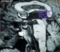
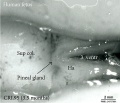
| Fetal Pineal Anatomy[5]
Superior (dorsal) view of the diencephalic-mesencephalic area of a 3.5-month-old human fetus. The third ventricle (3 ventr) without pial covering is seen to the right in the micrograph. The small pineal gland is a small protuberance (arrow) and merging via the broad stalk with the habenula (Ha). Sup col.: superior colliculus. Bar = 2 mm. |

|
Melatonin

- Melatonin is synthesized from the amino acid tryptophan within the pinealocytes.
- Serotonin is first acetylated by aryl alkylamine N-acetyltransferase (AA-NAT), then converted to melatonin by acetyl serotonin methyl transferase (ASMT also known as hydroxyindole O-methyltransferase or HIOMT).
- Melatonin release is stimulated by darkness and inhibited by light and is said to have neurological "chronobiotic" properties for resynchronization of sleep and circadian rhythms disturbances. In the periphery, melatonin is also involved in the regulation of several complex cycles: seasonal reproduction, body weight and energy balance.
- Melatonin levels can be monitored by urinary excretion of the melatonin metabolite 6-sulfatoxymelatonin (aMT.6S).
Melatonin Receptors
The hormone melatonin acts through receptors (high affinity G protein-coupled) embedded in the cell membrane. Three different receptor subtypes have been identified in mammals: MT1 (Mel 1a) and MT2 (Mel 1b) and a putative binding site called MT3.
- MT1 - expressed in humans in the pars tuberalis of the pituitary gland and the suprachiasmatic nuclei of the hypothalamus.
- MT2 - expressed in the retina.
- MT3 - expressed in many non-mammalian vertebrates in a range of brain areas.
Innervation
The gland is connected to the hypothalamus suprachiasmatic nucleus (SCN) central rhythm generator through a multi-synaptic pathway.
Nerve fibers innervating the mammalian pineal gland originate from perikarya located in the sympathetic superior cervical ganglion, the parasympathetic sphenopalatine and otic ganglia, as well as by nerve fibers originating in the central nervous system.[6]
- sympathetic nerves - contain norepinephrine and neuropeptide Y as neurotransmitters
- parasympathetic nerves - contain vasoactive intestinal peptide and peptide histidine isoleucine
- trigeminal ganglion - containing substance P, calcitonin gene-related peptide, and pituitary adenylate cyclase-activating peptide
Molecular Development
- Nodal - zebrafish required for dorsal convergence of pineal precursors.[7]
- Pax6 - rat pineal gland from E16, peak expression around E18.[8]
- Fgf8a - zebrafish epithalamus acts permissively to promote parapineal fate.[9]
- DARPP-32 (Dopamine- and cAMP-regulated phosphoprotein of 32 kDa) is involved in the retinal pathway transmitting photic information that resets the circadian clock.
Links: Molecular Development
Abnormalities
- Pineal Hypoplasia associated with retinal disease.
- Pineal Tumours in children are associated with abnormal puberty development.
Histology

Adult Histology
- Astrocytes - small dark nuclei
- Pinealocytes - most nuclei present, larger lighter and round nuclei surrounded by a broad rim of light cytoplasm
- Endothelial cells - nuclei in association with the vessels and capillaries traversing the tissue.
- Cytoplasmic processes - "stringy" appearance from both pinealocytes and astrocytes
- Links: large histology image
Images
Historic
| Historic Disclaimer - information about historic embryology pages |
|---|
|
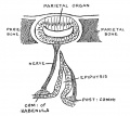
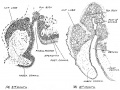
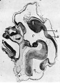
Cooper, ERA. The Human Pineal Gland and Pineal Cysts (1932)

|

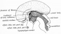
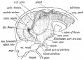
|
| Fig. 1. Sagittal midline section through head of 25 mm. embryo. x 8. A, anlage of pineal gland in the form of a backward hollowextension. B, anlage of posterior commissure. C, third ventricle. D, pituitary body. | Fig. 2. Sagittal section through head of 35 mm. embryo, not quite median. x 10. A, anterior anlage of pineal. B, posterior anlage with divertioulum pineale. C, posterior commisaure. D, third ventricle. E, pituitary body. |
References
Online Textbooks
- Pineal Gland and Cancer-An Epigenetic Approach to the Control of Malignancy: Evaluation of the Role of Melatonin Eurekah Bioscience Collection - Neuropharmacology
- Endocrine changes in puberty Endocrinology -> The gonad
- Second Malignancies Cancer Medicine -> Section 24: The Eye -> 85. Neoplasms of the Eye -> Pediatric Ophthalmic Oncology: Ocular Diseases
- The Action of Melatonin on Experimental in-Vivo Tumors Eurekah Bioscience Collection -> Neuropharmacology -> Pineal Gland and Cancer-An Epigenetic Approach to the Control of Malignancy: Evaluation of the Role of Melatonin -> Effect of Melatonin on Tumor Growth
- Potential Significance of (Patho)Physiological Changes of Melatonin for the Aetiology of Cancer Eurekah Bioscience Collection -> Neuropharmacology -> Pineal Gland and Cancer-An Epigenetic Approach to the Control of Malignancy: Evaluation of the Role of Melatonin
- Effects of Exogenous Melatonin AHRQ Evidence reports and summaries -> AHRQ Evidence Reports, Numbers 61 - 119 -> 108. Mel
Journals
- Journal of Pineal Research Molecular, Biological, Physiological and Clinical Aspects of Melatonin
Reviews
<pubmed></pubmed> <pubmed></pubmed> <pubmed></pubmed> <pubmed>16021838</pubmed> <pubmed>15589268</pubmed> <pubmed>15352385</pubmed> <pubmed>14561326</pubmed> <pubmed>9852259</pubmed> <pubmed></pubmed>
Articles
<pubmed>19665543</pubmed> <pubmed>16182388</pubmed> <pubmed>8938291</pubmed>
Search PubMed
Search April 2010
- Endocrine Development - All (14277) Review (4620) Free Full Text (3140)
Search Pubmed: pineal development
External Links
External Links Notice - The dynamic nature of the internet may mean that some of these listed links may no longer function. If the link no longer works search the web with the link text or name. Links to any external commercial sites are provided for information purposes only and should never be considered an endorsement. UNSW Embryology is provided as an educational resource with no clinical information or commercial affiliation.
- NIH The Julius Axelrod Papers | The Pineal Gland and the "Melatonin Hypothesis," 1959-1974
- NIH Child Health and Human Development (USA) Pineal Gland and Chronobiology: Regulation of Pineal Function
- University of Cincinnati SURVEY OF ENDOCRINE ORGANS
Additional Images
- Cooper1932-fig01.jpg File:Cooper1932-fig02.jpg

Historic Images
| Historic Disclaimer - information about historic embryology pages |
|---|
|
Keith1902 Fig 17
Keith1902 Fig 167
Keith1902 Fig 173
Keith1921 Fig 91
Keith1921 Fig 100
Keith1921 Fig 104
Keith1921 Fig 105
Cooper1932 Fig 1


Terms
Glossary Links
- Glossary: A | B | C | D | E | F | G | H | I | J | K | L | M | N | O | P | Q | R | S | T | U | V | W | X | Y | Z | Numbers | Symbols | Term Link
Cite this page: Hill, M.A. (2024, April 24) Embryology Endocrine - Pineal Development. Retrieved from https://embryology.med.unsw.edu.au/embryology/index.php/Endocrine_-_Pineal_Development
- © Dr Mark Hill 2024, UNSW Embryology ISBN: 978 0 7334 2609 4 - UNSW CRICOS Provider Code No. 00098G