Endocrine - Gonad Development: Difference between revisions
From Embryology
No edit summary |
|||
| Line 1: | Line 1: | ||
<div style="background:#F5FFFA; border: 1px solid #CEF2E0; padding: 1em; margin: auto; width: 90%; float:left;"><div style="margin:0;background-color:#cef2e0;font-family:sans-serif;font-size:120%;font-weight:bold;border:1px solid #a3bfb1;text-align:left;color:#000;padding-left:0.4em;padding-top:0.2em;padding-bottom:0.2em;">Notice - Mark Hill</div>Currently this page is only a template and will be updated (this notice removed when completed).</div> | |||
==Introduction== | ==Introduction== | ||
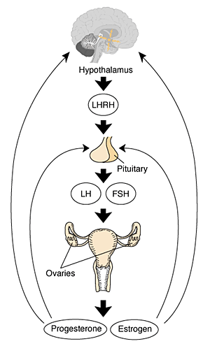
[[File:XXhpgaxis.gif|thumb|Female HPG axis]] | [[File:XXhpgaxis.gif|thumb|Female HPG axis]] | ||
Revision as of 18:40, 5 October 2010
Notice - Mark Hill
Currently this page is only a template and will be updated (this notice removed when completed).Introduction
Note this section of notes refers to the development of the gonad as an endocrine organ. A detailed description of the gonad development is covered in Ovary Development and Testis Development.
| Lecture - Genital Development | original page
HPG Axis - Endocrinology - Simplified diagram of the actions of gonadotrophins
Gonad Development
- mesoderm - mesothelium and underlying mesenchyme, primordial germ cells
- Gonadal ridge - mesothelium thickening, medial mesonephros
- Primordial Germ cells - yolk sac, to mesentery of hindgut, to genital ridge of developing kidney
Differentiation
- testis-determining factor (TDF) from Y chromosome: presence (testes), absence (ovaries)
Testis
- 8 Weeks, mesenchyme, interstitial cells (of Leydig) secrete testosterone, androstenedione
- 8 to 12 Weeks - hCG stimulates testosterone production
- Sustentacular cells - produce anti-mullerian hormone to puberty
Ovary
- X chromosome genes regulate ovary development
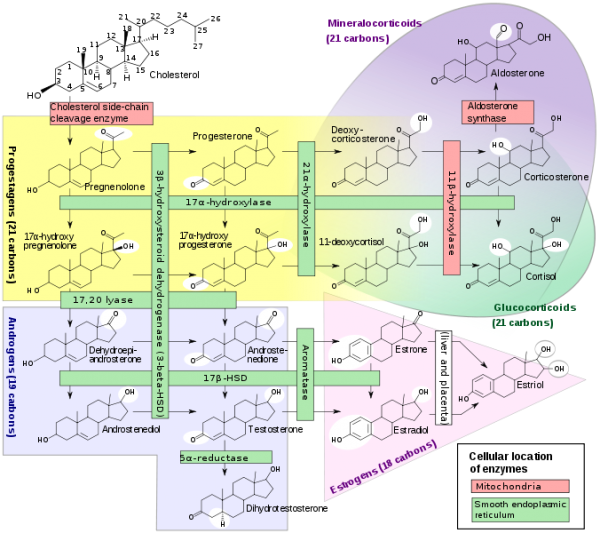
Steroidogenesis
References
Reviews
Articles
Search PubMed
Search Pubmed: endocrine gonad development
Additional Images
Adult Histology
Terms
Glossary Links
- Glossary: A | B | C | D | E | F | G | H | I | J | K | L | M | N | O | P | Q | R | S | T | U | V | W | X | Y | Z | Numbers | Symbols | Term Link
Cite this page: Hill, M.A. (2024, April 18) Embryology Endocrine - Gonad Development. Retrieved from https://embryology.med.unsw.edu.au/embryology/index.php/Endocrine_-_Gonad_Development
- © Dr Mark Hill 2024, UNSW Embryology ISBN: 978 0 7334 2609 4 - UNSW CRICOS Provider Code No. 00098G

