Embryonic Development: Difference between revisions
mNo edit summary |
mNo edit summary |
||
| Line 30: | Line 30: | ||
|} | |} | ||
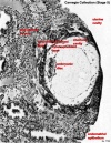
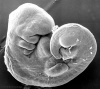
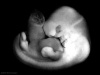
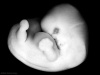
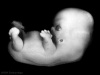
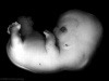
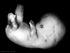
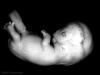
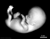
[[File:Stage14compare23.jpg|200px|right|Human Embryo]] | [[File:Stage14compare23.jpg|200px|right|Human Embryo]] | ||
| Line 52: | Line 43: | ||
Alternatively, look through development week by week. | Alternatively, look through development week by week. | ||
{{Carnegie_stages}} | |||
|} | |||
{{Week}} | {{Week}} | ||
== Week 1 == | == Week 1 == | ||
Revision as of 12:27, 12 June 2014
| Embryology - 23 Apr 2024 |
|---|
| Google Translate - select your language from the list shown below (this will open a new external page) |
|
العربية | català | 中文 | 中國傳統的 | français | Deutsche | עִברִית | हिंदी | bahasa Indonesia | italiano | 日本語 | 한국어 | မြန်မာ | Pilipino | Polskie | português | ਪੰਜਾਬੀ ਦੇ | Română | русский | Español | Swahili | Svensk | ไทย | Türkçe | اردو | ייִדיש | Tiếng Việt These external translations are automated and may not be accurate. (More? About Translations) |
Introduction


| Author Comments |
|---|
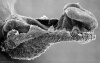
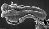
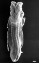
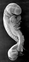
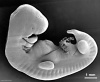
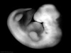
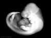
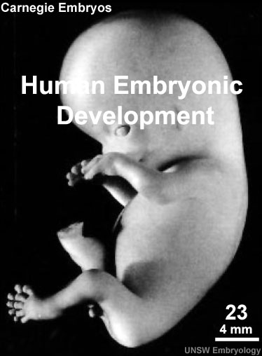
 Start here by looking at the external appearance of embryos in sequence from 1 to 23. Start here by looking at the external appearance of embryos in sequence from 1 to 23.
It is not so important to memorise the dates, as they are only approximate, but more important to understand growth (size changes) and the development (overall sequence of events) during this period.
|
| This page shows some key events of human development during the embryonic period of the first eight weeks (weeks 1 - 8) following fertilization. This period is also considered the organogenic period, when most organs within the embryo have begun to form.
|
|

| <mediaplayer width='374' height='525' image="http://php.med.unsw.edu.au/embryology/images/6/6e/Embryo_stages_003_icon.jpg">file:Embryo stages 003.mp4</mediaplayer> | Use the above stage number links to images and information about each specific stage of human development over the first 8 weeks. The links below give a broad overview of developmental events during each week.
Alternatively, look through development week by week. |
Embryo Week: Week 1 | Week 2 | Week 3 | Week 4 | Week 5 | Week 6 | Week 7 | Week 8 | Week 9
Week 1

- Week 1 Carnegie stage - 1 | 2 | 3 | 4
- Oocyte | Spermatozoa | Fertilization
- Zygote
- Morula
- Blastocyst
| Carnegie stages | |||
|---|---|---|---|
|
|
|
|
| stage 1 | stage 2 | stage 3 | stage 4 |
Week 2
- Week 2 Carnegie stage - 5 | 6
- Trophoblast - outer cell layer
- Embryoblast - inner cell mass
- Implantation
- Bilaminar embryo
| Carnegie stages | |
|---|---|
|
|
| stage 5 | stage 6 |
Week 3

| Carnegie stages | ||
|---|---|---|
|
|
|
| stage 7 | stage 8 | stage 9 |
Week 4

| Carnegie stages | |||
|---|---|---|---|
|
|
|
|
| stage 10 | stage 11 | stage 12 | stage 13 |
Week 5

| Carnegie stages | |
|---|---|
|
|
| stage 14 | stage 15 |
Week 6

| Carnegie stages | |
|---|---|
|
|
| stage 16 | stage 17 |
Week 7

| Carnegie stages | |
|---|---|
|
|
| stage 18 | stage 19 |
Week 8

| Carnegie stages | |||
|---|---|---|---|
|
|
|
|
| stage 20 | stage 21 | stage 22 | stage 23 |
- Carnegie Stages: 1 | 2 | 3 | 4 | 5 | 6 | 7 | 8 | 9 | 10 | 11 | 12 | 13 | 14 | 15 | 16 | 17 | 18 | 19 | 20 | 21 | 22 | 23 | About Stages | Timeline
Carnegie Stage Table
Weeks shown in the table below are embryonic post ovulation age, for clinical Gestational Age (GA) measured from last menstrual period, add 2 weeks.
(not to scale) |
||||
 |
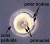
fertilized oocyte, zygote, pronuclei | |||
 |
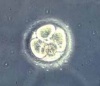
morula cell division with reduction in cytoplasmic volume, blastocyst formation of inner and outer cell mass | |||
 |
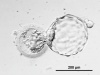
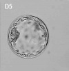
loss of zona pellucida, free blastocyst | |||
| attaching blastocyst | ||||
(week 2) |
 |
implantation | ||
 |
extraembryonic mesoderm, primitive streak, gastrulation | |||
| gastrulation, notochordal process | ||||
| primitive pit, notochordal canal | ||||
 |
Somitogenesis Somite Number 1 - 3 neural folds, cardiac primordium, head fold | |||
| Somite Number 4 - 12 neural fold fuses | ||||
| Somite Number 13 - 20 rostral neuropore closes | ||||
| Somite Number 21 - 29 caudal neuropore closes | ||||
| Somite Number 30 leg buds, lens placode, pharyngeal arches | ||||
| lens pit, optic cup | ||||
| lens vesicle, nasal pit, hand plate | ||||
| nasal pits moved ventrally, auricular hillocks, foot plate | ||||
| finger rays | ||||
| ossification commences | ||||
| straightening of trunk | ||||
| upper limbs longer and bent at elbow | ||||
| hands and feet turned inward | ||||
| eyelids, external ears | ||||
| rounded head, body and limbs | ||||
The embryos shown in the table are from the Kyoto and Carnegie collection and other sources.
Glossary Links
- Glossary: A | B | C | D | E | F | G | H | I | J | K | L | M | N | O | P | Q | R | S | T | U | V | W | X | Y | Z | Numbers | Symbols | Term Link
Cite this page: Hill, M.A. (2024, April 23) Embryology Embryonic Development. Retrieved from https://embryology.med.unsw.edu.au/embryology/index.php/Embryonic_Development
- © Dr Mark Hill 2024, UNSW Embryology ISBN: 978 0 7334 2609 4 - UNSW CRICOS Provider Code No. 00098G