Embryology History - William Smellie: Difference between revisions
| Line 490: | Line 490: | ||
Exhibits another front view of the Gravid Uterus in the beginning of labour ; the anterior parts being removed, as in the former Table ; but in this the Membranes, not being broken, form a large bag containing the Waters and Foetus. | Exhibits another front view of the Gravid Uterus in the beginning of labour ; the anterior parts being removed, as in the former Table ; but in this the Membranes, not being broken, form a large bag containing the Waters and Foetus. | ||
{| | |||
| [[File:Smellie1754 table_11.jpg|600px]] | |||
| '''Legend''' | |||
<gallery> | |||
A The substance of the uterus. | A The substance of the uterus. | ||
BCD The bones of the pelvis. | |||
E The coccyx. | E The coccyx. | ||
| Line 499: | Line 502: | ||
F The inferior part of the rectum. | F The inferior part of the rectum. | ||
G The vagina. | |||
H The mouth of the womb largely stretched in time of a pain ; with I, the membranes and waters. This circumstance makes it usually certain that labour is begun; whereas from the degree of dilatation represented in the former Table there is little to be ascertained, unless the pains are regular and strong, the os uteri being often found more open several days, and even weeks before labour commences. | |||
K The chorion. | K The chorion. | ||
| Line 508: | Line 511: | ||
M The placenta; the external convex surface of which, divided into a number of lobes, is here represented, its concave internal parts being covered by the chorion. | M The placenta; the external convex surface of which, divided into a number of lobes, is here represented, its concave internal parts being covered by the chorion. | ||
|} | |||
The placenta has been found adhering to all the different parts of the internal surface of the uterus, and sometimes even over the inside of the os uteri; this last manner of adhesion however always occasions noodings as soon as the same begins to dilate. | The placenta has been found adhering to all the different parts of the internal surface of the uterus, and sometimes even over the inside of the os uteri; this last manner of adhesion however always occasions noodings as soon as the same begins to dilate. | ||
| Line 518: | Line 523: | ||
Vide Vol. I. Book 3. Chap. 1. Sect. 4.. Chap. 2. Sect. 2, 5. Vol. II. Coll. 14, 23. | Vide Vol. I. Book 3. Chap. 1. Sect. 4.. Chap. 2. Sect. 2, 5. Vol. II. Coll. 14, 23. | ||
<gallery> | |||
File:Smellie1754 table_11.jpg| | |||
File:Smellie1754 table11.jpg| | |||
</gallery> | |||
==Twelfth Table== | ==Twelfth Table== | ||
Revision as of 08:24, 13 November 2012

William Smellie (1697-1763) A sett of anatomical tables, with explanations, and an abridgment, of the practice of midwifery (1754) He also helped develop the delivery forceps which by the late eighteenth century were a well-known standard obstetrical instrument.

Table 10 from A sett of anatomical tables, with explanations, and an abridgment, of the practice of midwifery (1754)
| Historic Disclaimer - information about historic embryology pages |
|---|
 |
A Set of Anatomical Tables with Explanations
And an Abridgement of the Practice of Midwifery; with a view to illustrate a treatise on that subject, and collection of cases by William Smellie M.D.
PHILADELPHIA:PRINTED BT A. BARTRAM, FOR THOMAS DOBSON, AT THE STONE HOUSE, NO. 41 y SOUTH SECOND STREET. 1806.
Preface
AS, in a long course of teaching and practice in Midwifery, I hope I may without vanity say, that I have done something towards reducing that Art into a more simple and mechanical method than has hitherto been done, I have attempted to explain the same in my Treatise of the Theory and Practice of Midwifery and Collection of Cases ; and finding that most of the representations hitherto given of the parts subservient to uterine gestation and parturition were in many respects deficient, I have been induced to undertake the following Tables, with a view to supply in some measure the defects of others, and at the same time to illustrate what I have taught and written on the subject. How far I have obtained those ends, it belongs to others to judge. I shall only beg leave to observe here by way of Preface, that the greatest part of the figures were taken from Subjects prepared on purpose, to shew every thing that might conduce to the improvement of the young Practitioner: Avoiding, however, the extreme minutia, and what else seemed foreign to the present design ; the situation of parts, and their respective dimensions, being more particularly attended to, than a minute anatomical investigation of their structure.
As these Tables may possibly fall into the hands of some who have not seen my former work, I have added an Abridgement of the Practice ; which, though far from being complete may serve to illustrate several things which otherwise, by a bare representation, would be hardly intelligible.
References are made to Vol. I. II. and III. By Vol. I. I mean that which I first published in the year 1752, and contains a view of the Theory and Practice of Midwifery ; Vol. II. and III. containing the Collection of Cases mentioned above. My first plan for these Tables confined them to the number of Twenty-two, which Mr. Eymsdyke had finished above two years ago ; but I soon saw that a further illustration, and consequently an addition to that number, was necessary. In eleven of these, Dr. Camper, formerly Professor of Medicine at Franequer in Friesland, now Professor of Anatomy and JBotany at Amsterdam^ greatly assisted me, viz. Table XII. XVI. XVII. XVIII. XIX. XXIV. XXVI. XXVII. XXVIII. XXXIV. and XXXVI The rest were drawn by Mr. Rymsdyke ; except the thirty-seventh and thirty-ninth, which were done by another hand. The whole of the drawings are faithfully engraved ; in which, however, delicacy and elegance have not been so much consulted as to have them done in a strong and distinct manner ; with this view chiefly, that from the cheap*ness of the work it may be rendered of more general use.
Explanations of a set of Anatomical Tables, with an Abridgement of the Practice of Midwifery (1754)

These historic tables below appear as they were republished in an 1806 textbook, preface shown above.
| Historic Disclaimer - information about historic embryology pages |
|---|
|
- Smellie 1754: Table 1 | Table 2 | Table 3 | Table 4 | Table 5 | Table 6 | Table 8 | Table 9 | Table 10 | Table 11 | Table 12 | Table 13 | Table 14 | Table 15 | All Tables
First Table
The First Table presents, in a front view, the Bones of a well-formed Pelvis.

|
Legend
A The five vertebra of the loins. B The os sacrum. C The os coccygis. D The ossa ilium. E The ossa ischium. F The ossa pubis. G The, foramina magna. H The acetabula. I The brim of the pelvis, or that circumference of its cavity, which is described at the sides of the inferior parts of the osssa ilium, and at the back and fore parts by the superior parts of the ossa pubis and sacrum. |

In this Table, besides the general structure and figure of the several bones, the dimensions of the brim of the pelvis, and the distance between the under parts of the ossa ischium, are particularly to be attended to ; from which it will appear, that the cavity of the brim is commonly wider from side to side than from the back to the fore part, but that the sides below are in the contrary proportion. The reader, however, ought not to conclude, that every pelvis is similar in figure and dimensions, since even well-formed ones differ in some degree from each other. In general, the brim of the pelvis measures about five inches and a quarter from side to side, and four inches and a quarter from the back to the fore part ; there being likewise the same distance between the inferior parts of the ossa ischium. All these measures, however, must be understood as taken from the skeleton; for, in the subject, the cavity of the pelvis is considerably diminished by its teguments and contents. Correspondent also to this diminution, the usual dimensions of the head of the full-grown foetus are but three inches and a half from ear to ear, and four inches and a quarter from the fore to the hind head.
Vide Tab. XVI. XVII. XVIII. Also Vol. I. Chap. I. Sect. 1. 2. 3. where the form and dimensions of the pelvis, as well as of the head of the fetus, and the manner in which the same is protruded in labour through the basin, are fully treated of. Consult likewise Vol. II. Coll. 1. No. 1. 2. where cases are given of complaints of the pelvis arising from difficult labours.
Second Table
Gives a lateral and internal view of the Pelvis divided longitudinally.

|
Legend
A The three lower vertebra? of the loins. B The os sacrum. C The os coccygis. D The left os ilium. E The left os ischium. F The os pubis of the same side. G The acute process of the os ischium. H The foramen magnum. I The brim of the pelvis. |

This Plate shews the distance from the superior part of the os sacrum to the ossa pubis, as well as from the last-mentioned bones to the coccyx, which in each amounts to about four inches and a quarter. The depth likewise of the posterior, lateral, and anterior parts of the pelvis, is shewn, not in the line of the body, but in that of the pelvis from its brim downward, which is generally three times deeper on the posterior than anterior part, and twice the depth of the last at the sides.
From this view appears also the angle which is formed by the last vertebra of the loins and the superior part of the os sacrum, as likewise the concavity or hollow space in the posterior internal part of the pelvis, arising from the curvature of the last-mentioned bone and coccyx ; finally, the distance from which to the posterior parts of the ossa ischium is here expressed.
Vide Tab. XVI. XVII. XVIII. XIX. Also Vol. I. and II as referred to in the former Table.
Third Table
Exhibits a front view of a distorted Pelvis.
| File:Smellie1754 table 03.jpg | Legend
A The five vertebra of the loins. B The os sacrum. C The os coccygis. D The ossa ilium. E The ossa ischium. F The ossa pubis. G The foramina magna. H The acetabula. |
From this Plate may appear the great danger incident to both mother and child when the pelvis is distorted in this manner ; it being only two inches and an half at the brim from the posterior to the anterior part, and the same distance between the inferior parts of each os ischium. Fide Tab. XXVII. where the pelvis is one quarter of an inch narrower at the brim than this, but sufficiently wide below. Various are the forms of distorted basins, but the last mentioned is the most common. It is a great happiness, however, in practice, that they are seldom so narrow, though there are instances where they have been much more so. The danger in all such cases must increase or diminish, according to the degree of distortion of the pelvis, and size of the child's head.
Fide Vol. I. Book I. Chap. 1. Sect= 4, 5. and Vol. IE Coll. 1. No. 3, 4, 5. Also Coll. 21, 27, and 29.
- Smellie1754 table03.jpg
Fourth Table
Shews the External Female Parts of Generation.
| File:Smellie1754 table 04.jpg | Legend
A The lower part of the abdomen. B The labia pudendi separated. C The clitoris and praputium. D The nymphte. E The fossa magna or os externum. F The meatus urinarius. G Thefrtznum labiorum. H The perineum. I The anus. K The part that covers the extremity of the coccyx. L The parts that cover the tuberosities of the ossa ischium. |
As it is of great consequence to every practitioner in midwifery, to know exactly the situation of the parts concerned in parturition, and which have not been accurately described by former anatomists with a view to this particular branch, I have given this draught from one of the preserved subjects which I keep by me, in order to demonstrate these parts in the ordinary course of my lectures. From a view, then of the situation of the parts, it appears, that the os externum is not placed in the middle of the inferior part of the pelvis, but at the anterior and inferior part of the pubes; and that the labia cover likewise the anterior part of these bones.
Secondly, It may be observed, that as the franum labiorum, which is nearly adjoining to the inferior part of the ossa pubis, is only about an inch from the anus, between which and the coccyx there is about three inches distance ; it follows, that the anus is nearer to the first mentioned bones than to the latter.
Thirdly, The view of this and the following Table will furnish proper hints with respect to the method of touching or examining the os uteri, without hurting or inflaming the parts; as it appears, that the os externum is placed forwards towards the pubes, and the os uteri backwards towards the rectum and coccyx. By this wise mechanism of nature many inconveniences are often prevented, which must happen if these parts were opposite to each other, and situated in the middle of the inferior part of the pelvis; particularly prolapsus of the vagina and uterus, either in the unimpregnated state, or in any of the first four months of pregnancy; as also too sudden deliveries in any of the last months.
Fourthly, From a view of the situation of the parts, it will appear, that in labour, when the os uteri is sufficiently opened to allow a passage for the head of the foetus, the same is protruded to the lower part of the vagina, by which the external parts are pushed out in form of a large tumour, as in Table XV.
Lastly, It may be observed, that when it is necessary to dilate the os externum, the principal force ought to be applied downwards and towards the rectum, to prevent the urethra and neck of the bladder from being hurt or inflamed.
Vide Vol. I. Book I. Chap. 2, Sect. 1. Vol. II. Coll. 2.
- Smellie1754 table 04.jpg
- Smellie1754 table04.jpg
Fifth Table
Figure I. Gives a front view of the Uterus in situ suspended in the vagina ; the anterior parts of the ossa ischium, with the 055a pubis, pudenda, perineum, and anus, being removed, in order to shew the internal parts.
| File:Smellie1754 table 05.jpg | Legend
B The ossa ilium. C The acetabula. D The inferior and posterior parts of the ossa ischium. Vide Table XXIX. where the ossa pubis and the anterior parts of the ossa ischium are represented by dotted lines. E The part covering the extremity of the coccyx. F The inferior part of the rectum. G The vagina cut open longitudinally, and stretched on each side of the collum uteri to shew in what manner the uterus is suspended in the same. FIH Part of the vesica urinaria stretched on each side of the vagina and inferior part of the fundus uteri. I The collum uteri. K The fundus uteri. L The tuba Fallopiana and fimbria. M The ovaria. N The ligamenta lata and rotunda. O The superior part of the rectum. |
Figure II. Gives a view of the internal parts 'as seen from the right groin, the pelvis being divided longitudinally.
A The lowest vertebra of the loins.
BC The os sacrum and coccyx, with the integuments.
D The left os ilium.
E The inferior part of the left os ischium.
F The os pubis of the same side.
G The foramen magnum.
H The acetabulum.
Ill The inferior part of the rectum and anus.
K The os externum and vagina ; the os uteri lying loosely in the same.
L The vesica urinaria.
MN The collum and fundus uteri, with a view of the cavity of both. The attachment of the vagina round the outside of the lips of the mouth of the womb is here likewise shewn, as also the situation of the uterus, as it is pressed downwards and backwards by the intestines and urinary bladder into the concave and inferior part of the os sacrum.
O The ligamenta lata and rotunda of the left side.
PP The Fallopian tube, with the fimbria ;
Q The ovarium of the same side.
RR The superior part of the rectum, and inferior part of the colon.
Figure III. Gives a front view of the UterUs in the beginning of the first month of pregnancy ; the anterior part being removed, that the Embryo might appear through the amnios, the chorion being dissected off.
A The fundus uteri.
B The collum uteri, with a view of the rugous canal that leads to the cavity of the fundus.
C The os uteri.
Vide Vol. I. Book I. Chap. 2. Sect. 2, 3. Vol. II. Coll. 3.
- Smellie1754 table 05.jpg
Sixth Table
Figure I. In the same view and section of the parts as in the first figure of the former table, shews the uterus as it appears in the second or third month of pregnancy, its anterior part being here likewise removed.

|
Legend
F The anus. G The vagina, with its plicae. H The posterior and inferior part of the urinary bladder extended on each side, the anterior and superior part being removed. I The mouth and neck of the womb, as raised up when examining the same by the touch, with one of the fingers in the vagina. K The uterus as stretched in the second or third month, containing the embryo, with the placenta adhering to the fundus. |

It appears from this and the former Table, that at this time nothing can be known, with respect to pregnancy, from the touch in the vagina, as the resistance of the uterus is so inconsiderable that it cannot prevent its being raised up before the finger ; and even were it kept down, the length of the neck would prevent the stretching being perceptible. The uterus likewise not being stretched above the pelvis, little change is made as to the figure of the abdomen, further than that the intestines are raised a little higher ; whence possibly the old observation of the abdomen being a little flatter at this period than usual, from the intestines being pressed more to each side. Women at this period miscarry oftener than at any other. It is a great happiness, however, in practice, that although they are frequently much weakened by large discharges, yet they rarely sink under the same, but are sooner or later relieved by labour coming on, which gradually stretches the neck and mouth of the womb, by the membranes being forced down with the waters ; and if the placenta is separated from the internal of the uterus, all its contents are discharged. But if Vat placenta still adheres, the membranes break, the waters and fatus are expelled, and the flooding diminishes, from the uterus, contracting close to the secundines, which also are usually discharged sooner or later.
From the structure, finally, of the parts, as represented in this and the former table, it may appear, that it is much safer to restrain the flooding, and support the patient, waiting with patience the efforts of nature, than to endeavour to stretch the os uteri, and deliver either with the hand or instruments, which might endanger a laceration and inflammation of the parts.
Vide C in Table XXXVII. Also Vol. I. Book II. Chap. 2. Sect. 2, 3, 4. Vol. II. Coll. 12. No. 2.

Figure II. Represents the uterus in the fourth or fifth month of pregnancy, in the same view and section of the parts with the former figure, excepting that in this the anterior part of the collum uteri is not removed.
In the natural situation, the mouth and lips of the womb are covered with the vagina, and these parts are contiguous to each other ; but here the vagina G is a little stretched from the neck and lips of the former, in order to shew the parts more distinctly. I, The neck of the womb, which appears in this figure thicker, shorter, and softer, than in the former. K, the inferior part of the fundus uteri ; the stretching of which can sometimes be felt through the vagina, by pushing up a finger on the anterior or lateral part of the same.
The uterus now is so largely stretched as to fill all the upper part of the pelvis, and begins also to increase so much as to rest on the brim, and to be supported by the same, the fundus at the same time being raised considerably above the pubes. From the abdomen being now more stretched the woman is more sensible of her growing bigger; and the uterus also, from the counter-pressure of the contents and parietes of the abdomen, is kept down, and the os uteri prevented from rising before the finger as formerly. In lean women, the stretching of the uterus can sometimes be perceived in the vagina at this period as well as above the pubes : But nothing certain can be discovered from the resistance or feel of the mouth of the womb or Ups y which are commonly the same in the first months of pregnancy as before it.
The size or bulk of the fetus is finally here to be observed, with the placenta adhering to the posterior part of the uterus.
Vide the references to Vol. I. and II- in the former Table.
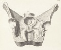
Table 6 Fig 1
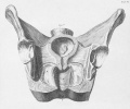
Table 6 Fig 1
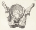
Table 6 Fig 2
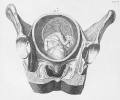
Table 6 Fig 2


Seventh Table
Represents the Abdomen of a woman opened in the sixth or seventh month of pregnancy. Figure I. In the same view and section of the parts as in the first figure of the former table, shews the uterus as it appears in the second or third month of pregnancy, its anterior part being here likewise removed.

|
Legend
A The parietes of the abdomen opened, and turned back to shew
|
The labia pudendi are sometimes affected in pregnancy with edematous swellings, occasioned by the pressure of the uterus upon the returning veins and lymphatics. If the labia are so tumefied as to obstruct the patient's walking, the complaint is removed by puncturing the parts affected. By which means the serous fluid is discharged for the present, but commonly incurs ; and the same operation must be repeated several times perhaps before delivery ; after which, however, the tumefaction entirely subsides. Here it may be observed, that this complaint can seldom or never obstruct delivery ; as the labia are situated at the anterior part of the ossapubis, and can rarely affect the stretching of the fr^num, perineum, vagina, and rectum. From this figure it appears, that the stretching of the uterus can easily be felt at this period in lean subjects, through the parietes of the abdomen ; especially if the intestines do not lie before it. In general indeed, as the uterus stretches, it rises higher ; by which means the intestines are likewise raised higher, and are also pressed to each side. Hence the nearer the woman is to her full time, the stretching is the more easily felt.
Vide Vol. I. Book I. Chap. 3. Sect. 3. Book III. Chap. l.Sect. 2. and Vol. II. Coll. 12, 13.

Eighth Table
In the same view and section of the parts as in Table VI. is represented the Uterus of the former Table, in order to shew its contents, and the internal parts as they appear in the sixth or seventh month of pregnancy.

|
Legend
A The uterus stretched up to the umbilical region. B The superior part of the ossa ilium. C The acetabula. D The remaining posterior parts of the ossa ischium. E The anus. F The vagina. G The bladder of urine. H The neck of the womb shorter than in Table VI. and raised higher by the stretching of the uterus above the brim of the pelvis. I The vessels of the uterus larger than in the unimpregnated state. K The placenta adhering to the inferior and posterior part of the uterus. L The membranes that surround the foetus. |
The fetal head of which is here represented (as well as of those in Table VI.) situated downwards at the inferior part of the uterus, and which I am apt to believe is the usual situation of the uterus when at rest and surrounded with a great quantity of waters, as the head is heavier than any other part. With respect to the situation of the body of the foetus, though the fore-parts are often turned towards the sides and posterior parts of the uterus, they are here, as well as in the foregoing Table, represented at the anterior part or fore wards, in order to shew them in a more distinct and picturesque manner.
Vide Vol. I. Book I. Chap. 3. Sect. 3, 4. Vol. II. Coll. 13. No. 1.
From this Table may appear the difficulty of stretching the os uteri in flooding cases, even at this period, from the length and thickness of the neck of the womb, especially in a first pregnancy : Much the same method, however, is to be followed here as was directed in Table VI. till labour comes on to dilate the os uteri. If the flooding is then considerable, the membranes should be broken, that the uterus may contract, and thereby lessen the discharge. The labour likewise, if it is necessary, may be assisted by dilating the os uteri in time of the pains ; which also, if wanting, may be provoked by the same method, when the patient is in danger. If this danger is imminent, and the woman seems ready to expire, the uterus, as appears from this Table, is at this time sufficiently stretched to receive the operator's hand to extract the fcetus, if the os internum can be safely dilated.
Lastly, It may be observed that women are in greater danger at this period and afterwards, than in the former months.
Vide Vol. I. Book III. Chap. 4. Sect. 3. No. 1, 2, 3. Vol. III. Coll. S3. No. 2. See also in the Edinburgh Physical and Literary Observations, Art. xvii. the dissection of a woman with child, by Dr. Donald Monro, Physician at London,
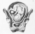
Table 8.

Ninth Table
In the same view and section of the parts with the former, represents the Uterus in the eighth or ninth month of pregnancy.

|
Legend
A The uterus as stretched to near its full extent, with the waters, and containing the foetus entangled in the funis, the head presenting at the upper part of the pelvis.
|
This and the foregoing Table shew in what manner the uterus stretches, and how its neck grows shorter, in the different periods of pregnancy ; as also the magnitude of the foetus, in order more fully to explain Vol. I. Book I. Chap. 3. Sect. 4, 5. also Lib. 3. Chap. 1. Sect. 1, 2. likewise Vol. II. Coll. 13. No. 1.
Notwithstanding it has been handed down as an invariable truth, from the earliest accounts of the art, to the present times, that, when the head of the foetus presented, the face was turned to the posterior part of the pelvis; yet, from Mr. Ould's observation, as well as from some late dissections of the gravid uterus, and what I myself have observed in practice, I am led to believe, that the head presents for the most part, as is here delineated, with one ear to the pubis, and the other to the os sacrum ; though sometimes this may vary, according to the form of the head, as well as that of the pelvis.
Consult Dr. Hunter's elegant plates of the gravid uterus.
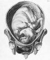
Table 8.

Tenth Table
Gives a front view of Twins in Utero in the beginning of labour ; the anterior parts being removed, as in the preceding Tables.

|
Legend
A The uterus as stretched with the membranes and waters. B The superior parts of the ossa ilium. C The acetabula. D The ossa ischium. E The coccyx. F The lower part of the rectum. G The vagina. H The os internum stretched open about a finger's breadth with the membranes and waters in time of labour-pains. I The inferior part of the uterus stretched with the waters which are below the head of the child that presents. K The two placentas adhering to the posterior part of the uterus, the two foetuses lying before them ; one with its head in a proper position, at the inferior part of the uterus; and the other situated preternaturally, with the head to the fundus: The bodies of each are here entangled in their proper funis, which frequently happens in the natural as well as preternatural positions. L The membranes belonging to each placenta. |
This representation of Twins, according to the order observed in my Treatise of Midwifery, ought to have been placed among the last Tables ; but, as that was of no consequence, I have placed it here in order to shew the os uteri grown much thinner than in the former figure, a little open and stretched by the waters and membranes which are pushed down before the head of one of the foetuses in time of a labourpain. With respect to the position of twins, it is often different in different cases ; but was thus, in a late dissection of a gravid uterus by Dr. Mackenzie.
Vide Vol. I. Book 3. Chap. 1. Sect. 4. and Chap. 5. Sect. 1. and Vol. II. Coll. 14. and Vol. III. Coll. 37.
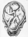
Table 10.
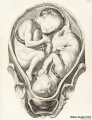
Table 10. Twins (small version)

Eleventh Table
Exhibits another front view of the Gravid Uterus in the beginning of labour ; the anterior parts being removed, as in the former Table ; but in this the Membranes, not being broken, form a large bag containing the Waters and Foetus.
| File:Smellie1754 table 11.jpg | Legend
Twelfth TableShews (in a lateral view and longitudinal division of the parts) the Gravid Uterus, when labour is somewhat advanced.
In this period of labour the os uteri being more and more stretched by the membranes pushing down, and beginning to extend the vagina, a great quantity of water is forced down at the same time, and (if the membranes break) is discharged ; whence the uterus contracts itself nearer to the body of iht fcetus, which is here represented in a natural position, with the vertex resting at the superior part of the ossa pubis, and the forehead towards the right os ilium. As soon as the uterus is in contact with the body of the fcetus, the head of the same is forced backward towards the os sacrum from the line of the abdomen EG into that of the pelvis, viz. from the uppermost F to near the end of the coccyx, and is gradually pushed lower as in the following Table. If the membranes do not break immediately upon their being pushed into the vagina, they should be allowed to protrude still further in order to dilate the os externum. Vide Vol. I. Book 1. Chap. 2. Sect. 2. Chap. 3. Sect. 3. Book 3. Chap. 1. Sect, l, 2. 4. Chap. 2. Sect. 3. Chap. 3. Sect. 4. No. 5. Vol. II. Coll. 10. No. 4. Case 3, 4. Coll. 14. Vol. III. Coll. 34. No. 2. Case 4.
Thirteenth TableIn the same view and section of the parts as in Table VI. shews the natural position of the head of the fetus when sunk down into the middle of the Pelvis after the Os Internum is fully opened, a large quantity of the waters being protruded with the Membranes through the Os Externum, but prevented from being all discharged, from the head's filling up the Vagina.
The vertex of the fatus being now down at the inferior part of the right os ischium, and the wide part of the head at the narrow and inferior part of the pelvis, the forehead by the force of the pains is gradually moved backwards ; and as it advances lower, the vertex and occiput turn out below the pubes, as in the next Table. Hence may be learnt of what consequence it is to know, that it is wider from side to side at the brim of the pelvis, than from the back to the fore part ; and that it is wider from the fore to the hind head of the child, than from ear to ear. Fide Vol. I. Book 1. Chap. I. Sect. 3, 5. Also Book 3. Chap. 3. Sect. 3, 4. No. 3. Vol. II. Coll. 14.
Fourteenth TableIn a similar view and section of the parts with Table XII.
Shews the forehead of the fetus turned (in its progression downwards, from its position in the former Table) backwards to the os sacrum and the occiput below the pubes ; by which means the narrow part of the head is to the narrow part of the pelvis, that is, between the inferior parts of the ossa ischium. Hence it may be observed, that though the distance between the inferior parts of the last mentioned bones is much the same as between the coccyx and pubes; yet as the cavity of the pelvis is much shallower at the anterior than lateral part, the occiput of the foetus, when come down to the inferior part of either os ischium, turns out below the pubes : this answers the same end as if the pelvis itself had been wider from the posterior part than from side to side ; the head likewise enlarging the cavity by forcing back the coccyx, and pushing out the external parts in form of a large tumour, as is more fully described in the following Table.
Vide Vol. I. 11. as referred to in the preceding Table. Fifteenth TableIs intended principally to shew in what manner the Perinaum and external parts are stretched by the head of the Foetus, in a first pregnancy, towards the end of labour. A The abdomen. W The labia pudendi. C The clitoris and its praputium. D The hairy scalp of the foetus swelled at the vertex in a laborious case, and protruded to the os externum. EF The perinaum and anus pushed out by the head of the foetus in form of a large tumour. GG The parts that cover the tuberosities of the ossa ischium. H The part that covers the os coccygis. The perinaum in this figure is stretched two inches, or double its length in the natural state, but when the os externum is so much dilated by the head of the foetus as to allow the delivery of the same, the perinaum is generally stretched to the length of three and sometimes four inches. The anus is likewise lengthened an inch, the parts also between it and the coccyx being much distended. Ail this ought to caution the young practitioner never to precipitate the delivery at this time; but to wait, and allow the parts to dilate in a slow manner ; as, from the violence of the labour-pains, the sudden delivery of the head of the fxtus might endanger the laceration of the parts. The palm of the operator's hand ought therefore to be pressed against the perinapum, that the head may be prevented from passing till the os externum is sufficiently dilated, to allow its delivery without tearing the framirn, and parts betwixt that and the a?ws, which are at this time very thin. Fide Vol. I. Book 3. Chap. 2. Sect. 2. Chap. 3. Sect. 4. No. 1. and Book 4. Chap. 1. Sect. 1. Also Vol. II. Coll. 14. 24. Vol. III. Coll. 40. Sixteenth TableAnd the three following shew in what manner the Head of the Foetus is helped along with the Forceps, as artificial hands, when it is necessary to assist with the same for the safety of either Mother or Child. In this Table the head is represented as forced down into the Pelvis by the labour-pains, from its former position in Table XII. AABC The vertebrae of the loins, os sacrum, and coccyx. D The os pubis of the left side. E The remaining part of the bladder. FF The intestinum rectum. GGG The uterus. H The mons veneris. I The clitoris, with the left nympha. X The corpus cavernosum clitoridis. V The meatus urinarius. K The left labium pudendi. L The anus. N The perineum. QP The left hip and thigh. R The skin and muscular part of the loins. The patient in this case may be, as in this Table, on her side, with her breech a little over the side or foot of the bed, her knees being likewise pulled up to her belly, and a pillow placed between them, care being taken at the same time that the parts are by a proper covering defended from the external air. If the hairy scalp of the fcetas is so swelled that the situation of the head cannot be distinguished by .the sutures as in Table XXI. or if by introducing a finger between the head of the child and the pubes, or groins, the ear or back part of the neck cannot be felt, the os externum must be gradually dilated in the time of the pains with the operator's fingers (previously lubricated with hog's-lard) till the whole hand can be introduced into the vagina, and slipped up in a flattish form between the posterior part of the pelvis and child's head. This last is then to be raised up as high as is possible, to allow room for the fingers to reach the ear and posterior part of the neck. When the position of the head is known, the operator must withdraw his hand, and wait to see if the stretching of the parts will renew or increase the labourpains, and allow more space for the advancement of the head in the pelvis. If this, however, proves of no effect, the fingers are again to be introduced as before, and one of the blades of the forceps (lubricated with lard) is then to be applied along the inside of the hand or fingers, and left ear of the child, as represented in the Table. But if the pelvis is distorted and projects forward at the superior part of the os sacrum, and the forehead therefore cannot be moved a little backwards, in order to turn the ear from that part of the pelvis which prevents the end of the forceps to pass the same ; in that case, I say, the blade must be introduced along the posterior part of the ear at the side of the distorted bone. The hand that was introduced is then to be withdrawn, and the handle of the introduced blade held with it as far back as the perinaum will allow, whilst the fingers of the other hand are introduced to the os uteri, at the pubes, or right groin, and the other blade placed exactly opposite to the former. This done, the handles being taken hold of and joined together, the head is to be pulled lower and lower every pain, till the vertex, as in this Table, is brought down to the inferior part of the left ischium, or below the same. The wide part of the head being now advanced to the narrow part of the pelvis betwixt the tuberosities of the ossa ischium, it is to be turned from the left ischium out below the pubes, and the forehead backwards to the concave part of the os sacrum and coccyx, as in Table XVII. and afterwards the head brought along and delivered as in Table XVIII. and XIX. But if it is found that the delivery will require a considerable degree of force from the head's being large, or the pelvis narrow, the handles of the forceps are to be tied together with a fillet, as represented in this Table, to prevent their position being changed, whilst the woman is turned on her back, as in Table XXIV which is then more convenient for delivering the head than when lying on the side. This Table shews that the handles of the forceps ought to be held as far back as the os externum will allow, that the blades may be in an imaginary line between that, and the middle space between the umbilicus and the scrobiculus cordis. When the. forceps are applied along the ears and sides of the head, they are nearer to one another, have a better hold, and mark less than when over the occipital and frontal bones. Vide Vol. I. Book 3. Chap. 3. from Sect. 1. to 6. and Vol. II. Coll. 25, 26, 27, and 29. Seventeenth TableIn the same view with the former, represents in outlines the Head of the Foetus brought lower with the Forceps, and turned from the position in the former Table, in imitation of the natural progression by the labour-pains, which may likewise be supposed to have made this turn, before it was necessary to assist with the Forceps, this necessity at last arising from many of the causes mentioned in Vol. I. In this view the position of the forceps, along the ears and narrow part of the head, is more particularly expressed. It appears also, that when the vertex is turned from the left os ischium, where it was closely confined, it is disengaged by coming out below the pubes, and the forehead that was pressed against the middle of the right os ischium is turned into the concavity of the os sacrum and coccyx. By this means the narrow part of the head is now between the ossa ischium or narrow part of the pelvis ; and as the occiput comes out below the pubes, the head passes still easier along. When the head is advanced so low in the pelvis, if the position cannot be distinguished by the sutures, it may for the most part be known by feeling for the back part of the neck of the foetus, with a finger introduced betwixt the occiput and pubes, or towards one of the grows. If the head is squeezed into a longish form, as in Table XXI. and has been detained many hours in this position the pains not being sufficient to complete the delivery, the assistance of the forceps must be taken to save the child, though the woman may be in no danger. But if the head is high up in the pelvis, as in the former Table, the forceps ought not to be used except in the most urgent necessity. This Table also shews that the handles of the forceps are still to be kept back to the perineum, and when in this position are in a line with the upper part of the sacrum, and if held more backwards, when the head is a little higher, would be in a line with the scrobiculus cordis. If the forceps are applied when the head is in this position, they are more easily introduced when the patient is in a supine position, as in Table XXIV. Neither is it necessary to tie the handles, which is only done to prevent their alteration when turning the woman from her side to her back. As 1 have had several cases where a longer sort of forceps that are curved upwards are of great use to help along the head, when the body is delivered first, as in Table XXXV. the same are represented here by dotted lines. They may be used in laborious cases as well as the others, but are not managed with the same ease.
LM The anus MN The perineum. O The common integuments of the abdomen. R The short forceps. S The long curved forceps. The first of these is eleven inches long, and the last twelve inches and a half, which I have after several alterations found sufficient ; but this need not confine others who may choose to alter them from this standard. Vide Table XXXVII. Eighteenth TableIn the same view and section of the parts, shews the Head of the Foetus in the same position, but brought lower down with the Forceps than in the former Table ; for in this the Os Externum is more open, the Occiput come lower down from below the Pubes, and the Forehead past the Coccyx, by which both the Anus and Perinaum are stretched out in form of a large tumour, as in Table XX.
abch The outlines of the os ilium. Def The same of the pubis and ischium* iik The acetabulum. And mn Thejbra?nen magnum. Vide Vol. I. Book 3. Chap. 5, Sect. 3. Vol. II. Coll. 25. Nineteenth TableIn the same view and section of the pelvis, is intended by outlines to shew, that as the external parts are stretched, and the os externum is dilated, the occiput of the foetus, rises up with a semicircular turn from out below the pubes, the under part of which bones are as an axis, or fulcrum, on which the back part of the neck turns, whilst at the same time the forehead and face, in their turn upwards, distend largely the parts between the coccyx and os externum. This is the method observed by nature in stretching these parts in labour; and as nature is always to be imitated, the same method ought to be followed when it is necessary to help along the head with the forceps. Vide the three former Tables for the descriptions and references. Twentieth TableIn the same section of the parts, but with a view of the right side, shews the Head of the Foetus in the contrary position to the three last figures, the Vertex being here in the concavity of the Sacrum, and tne Forehead turned to the Pubes. AB The vertebra of the loins, os sacrum, and coccyx. C The os pubis of the right side. D The anus. E The os externum not yet begun to stretch. F The nympha. G The labium pudendi of the right side. H The hip and thigh. II The uterus contracted, the waters being all discharged. When the head is small, and the pelvis large, the parietal bones and the forehead will, in this case, as they are forced downwards by the labour-pains, gradually dilate the os externum, and stretch the parts between that and the coccyx in form of a large tumour, as in Table XV. till the face comes down below the pubes, when the head will be safely delivered. But if the same be large, and the pelvis narrow, the difficulty will be greater, and the child in danger ; as in the following Table. Vide Vol. I. Book 3. Chap. 3. Sect. 4. No. 3. Vol. II. Coll. 16. No. 2. Twenty-First TableShews the head of the Foetus in the same position as in the former Table ; but, being much larger, it is by strong labour-pains squeezed into a longish form with a Tumour on the Vertex, from the long compression of the head in the Pelvis. If the Child cannot be delivered with the labour-pains, or turned and brought footling, the Forceps are to be applied on the head, as described in this figure-, and brought along as it presents ; but if that cannot be done without running the risk of tearing the Perineum, and even the Vagina and Rectum of the Woman, the Forehead must be turned backwards to the Sacrum. To do this more effectually, the Operator must grasp firmly with both hands the handles of the Forceps, and at the same time pushing upwards raise the Head as high as possible, in order to turn the Forehead to one side, by which it is brought into the natural position ; this done, the head may be brought down and delivered as in Table XVI. he. Vide Vol. I. Book 3. Chap. 3. Sect. 4. No. 2. and Vol. II. Coll. 28. Also the former Table for the description of the parts, except K The tumour on the vertex. The same compression and elongation of the head, as well as the tumour on the vertex, may be supposed to happen in a greater or less degree in the XVI. XVII. XVIII. XIX. Tables, as well as in this, where the difficulty proceeds from the head being large, or the pelvis narrow. Vide Table XXVII. and XXVIII. Sometimes the forehead maybe moved to the natural position by the assistance of the fingers, or only one blade of the forceps. The forceps may either be the straight kind, or such as are curved to one side, when it is necessary to use one or both blades. M The vesica urinaria much distended with a large quantity of urine from the long pressure of the head against the urethra; which shews that the urine ought to be drawn off with a catheter, in such extraordinary cases, before you apply the forceps or in preternatural cases where the child is brought footling. N The under part of the uterus. 00 The os uteri. Twenty-Second TableShews in a front view of the parts, the Forehead of the Foetus presenting at the brim of the Pelvis ; the Face being turned to one side ; the Fontanelle to the other ; and the Feet and Breech stretched towards the Fundus Uteri. AA The superior part of the ossa ilium. B The anus. C The perineum. D The os externum; the thickness of the posterior part before it is stretched with the head of the child. EEE The vagina. F The os uteri, not yet fully dilated. GGG The uterus. H The membrana adiposa. If the face is not forced down, the head will sometimes come along in this manner, in which case the vertex will be flattened, and the forehead raised in a conical form ; and when the head comes down to the lower part of the pelvis, the face or occiput will be turned from the side, and come out below the pubes. But if the head is large and cannot be delivered by the pains, or if the wrong position cannot be altered, the child must, if possible, be brought footling, or delivered with the forceps. Vide Vol. I. Book 3. Chap. 2. Sect. 3. Chap. 3. Sect. 4. No. 3. Vol. II. Coll. 16. No. 4. Coll. 28. Twenty-Third TableShews, in a lateral view, the Face of the Child presenting, and forced down into the lower part of the Pelvis, the Chin being below the Pubes, and the Vertex in the concavity of the Os Sacrum : The waters likewise being all discharged, the Uterus appears closely joined to the body of the Child, round the neck of which is one circumvolution of the Funis. AB The vertebra of the loins, os sacrum, and coccyx. C The os pubis of the left side. D The inferior part of the rectum. E The perinaum. F The left labium pudendi. GGG The vterus. When the pelvis is large, the head, if small, will come along in this position, and the child be saved : For as the head advances lower, the face and forehead will stretch the parts between the franum labiorum and coccyx in form of a large tumour. As the os externum likewise is dilated, the face will be forced through it ; the under part of the chin will rise upwards over the anterior part of the pubes ; and the forehead, vertex, and occiput, turn up from the parts below. If the head, however, is large, it will be detained either when higher or in this position. In this case, if the position cannot be altered to the natural, the child ought to be turned, and delivered footling. If the pelvis, however, is narrow, and the waters not all gone, the vertex should, if possible, be brought to present; but if the uterus is so closely contracted that this cannot be effected, on account of the strong pressure of the same, and slipperiness of the child's head, in this case the method directed in the following Table is to be taken. Twenty-Fourth TableRepresents, in the lateral view, the Head of the Foetus, in the same position as in the former Table ; but the delivery is supposed to be retarded from the largeness of the Head, or a narrow Pelvis.
Twenty-Fifth TableShews, in a lateral view of the right side, the Face of the Foetus presenting, as in Table XXlil. but in the contrary position; that is with the chin to the Os Sacrum, and the Bregma to the Pubes, the Waters evacuated, and the Uterus contracted. A The os externum not yet begun to stretch; B The anus. Fide Table XX. for the further description of the parts.
Twenty-Sixth TableRepresents by outlines, in a lateral view of the left side of the subject, the Foetus in the same situation as in the former Table.
Twenty-Seventh TableGives a lateral internal view of a distorted Pelvis, divided longitudinally, with the Head of a Fcetus of the seventh month passing the same. Vide the Explanation of Table III.
Twenty-Eighth TableGives a side-view of a distorted Pelvis, as in the former Table, with the Head of a fullgrown Fa?tus, squeezed into the Brim, the Parietal Bones decussating each other, and compressed into a conical form.
Twenty-Ninth TableRepresents, in a front view of the Pelvis, as in Table XXII. the Breech of the Foetus presenting, and dilating the Os Internum, the Membranes being too soon broke. The foreparts of the Child are to the posterior part of the Uterus ; and the Funis with a knot upon it surrounds the neck, arm, and body.
Vide Vol. I. Book 3. Chap. 4. Sect. 1, 2. Vol. III. Coll. 32. The description of the parts in this, and the following Table, is the same as in Table XXII ; only the dotted lines in this describe the place of the ossa pubis, and anterior parts of the ossa ischium which are removed, and may serve in this respect as an example for all the other frontviews, where, without disfiguring the Table, they could not be so well put in.
Thirtieth TableShews, in the same view and with the same references as the former, the breech oi'ihefatus presenting; with this difference, however, that the fore-parts of the child are to the fore-part of the uterus. In this case, when the breech coming double as it presents is brought down to the hams, the legs must be extracted, a cloth wrapped round them, and the fore-parts of the child turned to the back-parts of the woman. If a pain should in the mean time force down the body of the child, it ought to be pushed up again in turning, as it turns easier when the belly is in the pelvis, than when the breast and shoulders are engaged ; and as sometimes the face and forehead are rather towards one of the groins, a quarter turn more brings these parts to the side of the pelvis, and a little backwards, after which the body is to be brought down. If the child is not large, the arms need not be brought down, and the head may be delivered by pressing back the shoulders and body of the child to the perina-um, and whilst the chin and face are within the vagina, to bring the occiput out from below the pubes, according to Daventer s method. Or the operator may introduce a finger or two into the mouth, or on each side of the nose, and, supporting the body on the same arm, fix two fingers of the other hand over the shoulders, on each side of the child's neck, and in this manner raise the body over thepubes, and bring the face and forehead out with a semicircular turn upwards, from the under part of the os externum. All this maybe easily done when the woman lies on her side ; but if the child is large, and the pelvis narrow, it is better to turn the patient on her back, as described in Table XXIV. ; and after the It- gs and body are extracted as far as the shoulders, the arms are to be cautiously brought down and the head delivered. If the woman has strong pains, and when by the felt pulsation of they uni "s umbilicalis, or the struggling motions of the fatus, it is certain that the child is still alive, wait with patience for the assistance of the labour : But if that and the hand are insufficient, and the pulsation of the funis turns weaker, and if the child cannot be brought double, the breech must be pushed up ; and if the resistance of the uterus is so great, as to prevent the extraction of the legs, the patient ought to be turned on her knees and elbows. When the legs are thus brought down, the woman, if needful, is to be again turned to her back, to allow more freedom to deliver the body and head, as before described. If the head after several trials cannot be delivered, without endangering the child, from overstraining the neck, the long curved forceps ought to be applied, as in Table XXXV. If these fail, and the patient is not in danger, some time may be allowed for the effect of the labour-pains ; which likewise proving insufficient, the crotchet must be used as in Table XXXIX. and when it is certain that the child is dead, or that there is no possibility of saving it. Thirty-First TableRepresents, in a front view of the Pelvis, the Foetus compressed, by the contraction of the Uterus into a round form, the fore-parts of the former being towards the inferior part of the latter, and one Foot and Hand fallen down into the Vagina. In this figure the anterior part of the Pelvis is removed by a longitudinal section through the middle of the Foramen Magnum. AA The superior parts of the ossa ilium. BB The uterus*. GG The mouth of the womb stretched, and appearing in OOOO The vagina. D The inferior and posterior part of the os externum. EEEE The remaining part of the ossa pubis and ischium. FFFF The membrana adiposa. This and the three following Tables, representing four different preternatural positions of xhtfatus in utero, may serve as examples for the manner of delivery in these as well as hi all other preternatural cases. In all preternatural cases, the foetus may be easily turned and delivered by the feet, if known before the membranes are broke, and the waters, discharged ; or if the pelvis is narrow, and the patient is strong, the head, if large, may be brought down so as to present in the natural way : but if all the waters are discharged, and the uterus is strongly contracted to the body of the fetus, this last method can seldom take place on account of the strong pressure of the uterus, and slipperiness of the child's head. In the present case, the woman may either be laid on her back or side, as described in Tables XVI. and XXIV. ; and the operator, having slowly dilated the os externum with his fingers, must introduce the same into the vagina, and push up into the uterus the parts of the fetus that present; or if there is space for it, his hand may pass in order to dilate the os internum if not sufficiently previously stretched by the membranes and waters. This done, he must advance his hand into the uterus to know the position of the foetus : and, as the breech is rather lower than the head, search for the other leg, and bring down both feet without the os externum. A cloth must then be wrapped round them ; and, having grasped them with one hand, he is to introduce the other into the uterus, in order to raise the head of the foetus, whilst the legs and thighs are pulled down by the hand that holds the feet. When the head is raised, and does not fall down again, the hand of the operator may be withdrawn from the uterus, and the delivery completed as directed in the two former Tables. By the artless method of taking hold and pulling one or both feet, the breech may come down and the head rise to the fundus ; but if this should not happen, there will be great danger of overstraining the foetus ; which is prevented by the former method. If the membranes are broken before the os uteri is largely opened, and the hand of the operator cannot be introduced, which sometimes happens in a first pregnancy, the parts of the foetus' should be allowed to protrude still further, by which means the rigidity of the os internum will in time be lessened. Vide Vol. I. and III. on preternatural labours. Thirty-Second TableRepresents, in the same view with the former, the Fcetus in the contrary Position ; the Breech and Fore-parts being towards the Fundus Uteri, the left Arm in the Vagina, and fore Arm without the Os Externum, the shoulder being likewise forced into the Os Uteri.
Fide the explanations and references of the foregoing Table to illustrate this and the following.
Thirty-Third TableExhibits, in the same view likewise of the Pelvis with the former, a third position of the Fatus when compressed into the round form ; the Belly, or Umbilical Region, presenting at the Os Internum, and the Funis fallen down into the Vagina, and appearing at the Os Externum.
Thirty-Fourth TableShews in a lateral view of the Pelvis, one of the most difficult preternatural cases. The left Shoulder, Breast, and Neck of the Faults presenting, the Head reflected over the Pubes to the right Shoulder and Back, and the Feet and Breech stretched up to the Fundus, the Uterus contracted at the same time, in form of a long Sheath, round the Body of the Foetus. ABC The os sacrum and coccyx. D The os pubis of the left side. E Part of the urinary bladder. F The rectum. KL The private parts. M The anus. MN The per in a nm. V The meatus urinarius. O The os uteri, not yet opened, and situated backwards towards the rectum and coccyx. RS The same represented in dotted lines, as opened when the labour is begun. IH The same more fully dilated, but nearer to the posterior than anterior part of the pelvis. WP The same not fully stretched at the forepart, though entirely obliterated at the backpart, the uterus and vagina being there only sometimes one continued surface.
Vide Vol. I. and III. as directed in Table XXXI. Thirty-Fifth TableShews in a lateral view of the Pelvis, the method of assisting the Delivery of the Head of the Fcetus with the long curved Forceps, in preternatural Cases, when it cannot be done with the hands as described in Tables XXIX. and XXX.
B The os pubis of the left side. CC The perinaum and anus pressed backwards with the forceps. D The. intestines. EE The parietes of the abdomen. FFF The uterus. G The posterior part of the os uteri. H The rectum. I The vagina. After the body and arms of the child are delivered, and the different methods used to bring down the head with the hands, as directed in the above Table, and more fully described in Vol. I. and III. the following method is to be tried in order to save the child, which must otherwise be lost by overstraining the neck and spinal marrow. The woman being in the supine position, as in Table XXIV. one of the assistants ought to hold the body and arms of the child up towards the abdomen of the woman, to give more room to the operator, who having introduced one hand up to the child's face, and moved it from the side a little backwards, for the easier application of the forceps along the sides of the head, must then turn his hand to one of the ears, and introduce one of the blades with the other hand between the same and the head, with the curved side towards the pubes, as in this Table. This done, the hand is to be brought down to hold the handle of the blade of the forceps, till the other hand is introduced to the other side of the head, by which means the same is pressed against the blade that is up, and which is thus prevented from slipping, whilst the other hand introduces the second blade on the opposite side. The blades being thus introduced, care must be taken, that in joining them no part of the vagina is locked in. After the forceps are firmly fixed along the sides of the head, the face and forehead must be turned again to the side of the brim of the pelvis, by which means the wide part of the head is to the wide part of the brim. This done, the head is to be brought lower, and the force gradually increased, according to the resistance from the largeness of the head, or narrowness of the pelvis. The forehead, when brought low enough down, is then to be turned into the concavity of the os sacrum and coccyx, the handles of the forceps raised upwards, and the same caution used in bringing the head through the os externum, as described in Table XiX. and XXX. By this method the head will be delivered, the child frequently saved, and the use of the crotchet prevented, except in those basins that are so narrow, that it is impossible to deliver without diminishing the bulk of the head. Vide Table XXXIX. Also Vol. I. Book 3. Chap. 4. Sect. 5. Vol. III. Coll. 34, 35. Thirty-Sixth TableRepresents, in a lateral view of the Pelvis, the method of extracting, with the assistance of a curved Crotchet, the head of the Foetus, when left in the Uterus, after the body is delivered and separated from it, either by its being too large, or the Pelvis too narrow. ABC The os sacrum and coccyx, D The os pubis of the left side. EE The uterus. F The locking part of the crotchet, ghi The point of the crotchet on the inside of the cranium.
Vide Vol. I. Book 3. Chap. 3. Sect. 7. Chap. 4. Sect. 5. Also Vol. III. Coll. 31, 36. Thirty-Seventh TableAnd the two following, represent several kinds of Instruments useful in laborious and difficult Cases. a The straight short forceps, in the exact proportion as to the width between the blades, and length from the points to the locking part: The first being two and the second six inches, which with five inches and a half (the length of the handles), makes in all eleven inches and a half. The length of the handles may be altered at pleasure. 1 find, however, in practice, that this standard is the most convenient, and with less difficulty introduced, than when longer, having also sufficient force to deliver in most cases where their assistance is necessary. The handles and lowest part of the blades may as here be covered with any durable leather ; but the blades ought to be wrapped round with something of a thinner kind, which may be easily renewed when there is the least suspicion of venereal infection in a former case : By being thus covered, the forceps can have a better hold, and mark less the head of the child. For their easier introduction, the blades ought likewise to be greased with hog's lard. b Represents the posterior part of a single blade in order to shew the open part of the same, and the form and proportion of the whole. The handles, however, as here represented, are rather too large. Vide Table XXI. for the figure and proportions of the long forceps, that are curved upwards, and covered in the same manner as the former. The forceps were at first contrived to save the fatus, and prevent, as much as possible the use of sharp instruments ; but even to this salutary method, recourse ought not to be had but in cases where the degree of force requisite to extract will not endanger, by its consequences, the life of the mother : For, by the imprudent use of the forceps, much more harm may be done than good. See the explanation of Table XVI. Also the preface to Vol. II. with the cases in the Collection on that subject. c The blunt hook which is used for three purposes. First, To assist the extraction of the head after the cranium is opened with the scissars, by introducing the small end along the ear on the out side of the head to above the under jaw. where the point is to be fixed ; the other extremity of the hook being held with one hand, whilst two fingers of the other are to be introduced into the aforesaid opening, by which holds the head is to be gradually extracted. Secondly, The small end is useful in abortions in any of the first four or five months, to hook down the secundines when lying loose in the uterus, when the patient is much weakened by floodings from the too long retention of the same, the pains also being unable to expel them, and when they cannot be extracted with the fingers. But i£ the placefita still adheres, it is dangerous to use this or any other instrument to extract the same, as it ought to be left till it separates naturally. If a small part of the secundines is protruded through the os uteri, and pulled away from what still adheres in the uterus, the mouth of the womb contracts, and that irritation is thereby removed which would have continued the pains, and have separated and discharged the whole. Thirdly, The large hook at the other end is useful to assist the extraction of the body, when the breech presents ; but should be used with great caution, to avoid the dislocation or fracture of the thigh. Vide Table XXIX. Also Vol. I. Book 2. Chap. 3. Book 3. Chap. 3. Sect. 7. and Chap. 4. Sect. 2. Vol. II. Coll. 12. Vol. III. Coll. 31. 32. Thirty-Eighth TableA, represents the whale-bone fillet, which may be sometimes useful in laborious cases, when the operator is not provided with the forceps in sudden and unexpected exigencies.
the the pubes, it requires one of this length ; and in some extraordinary cases, I have been obliged to use a male catheter. Vide Vol. I. Book 2. Chap. 1. Sect. 1, 2. Vol. II. Coll. 10. No. 2. Thirty-Ninth Tablea Represents a pair of curved crotchets locked together in the same manner as the forceps. It is very rare that the use of both is necessary, excepting when the face presents with the chin turned to the sacrum, and when it is impossible to move the head to bring the child footling, or deliver with the forceps. In that case, if one crotchet is not sufficient, the other is to be introduced, and when joined together will act both as crotchets, in opening the cranium, and, as the head advances, will likewise act as forceps in moving and turning the head more conveniently for the delivery of the same. They may also be useful to assist when the head is left in the uterus, and one blade is not sufficient. There is seldom occasion, however, for the sharp crotchet, when the head presents ; the blunt hook in Table XXVII. being commonly sufficient, or even the forceps to extract the same, after it is opened with the scissers. Great care ought to be taken, when the sharp crotchet is introduced, to keep the point towards the fetus, especially in cases where the fingers cannot be got up to guide the same. The dotted lines along the inside of one of the blades, represent a sheath that is contrived to guard the point till it is introduced high enough ; the ligature at the handles marked with the two dotted lines is then to be untied, tied, the sheath withdrawn, and the point being uncovered, is fixed as directed in Table XXXVI. The point, guarded with this sheath, may also be used instead of the blunt hook. b Gives a view of the back-part of one of the crotchets, which is twelve inches long. c Gives a front-view of the point, to shew its length and breadth, which ought to be rather longer and narrower than here represented. d Represents the scissars proper for perforating the cranium in very narrow and distorted pehises. They ought to be made very strong, and nine inches at least in length, with stops or rests in the middle of the blades, by which a large dilatation is more easily made. The above instruments ought only to be used in the most extraordinary cases, where it is not possible to save the woman without their assistance. Fide Vol. I. Book 3. Chap. 3. Sect. 5. Chap. 5. No. 1. Vol. III. Coll. 31, 3 5.
Additional TableNumb. XL. Among the few improvements which have been made in the obstetrical apparatus since the days of Dr. Smellie, the most important are the alterations in the forceps, by which the inconveniencies formerly attending the use of that instrument are obviated, and the operation is rendered more safe and easy. In contriving these alterations, the intentions were, 1. That the large curves should correspond as nearly as possible with that of the pelvis. 2. That their points should be thrown forwards, and made round, to prevent their hitching, or even pressing uneasily against any part of the pelvis; and likewise to maintain their hold of the head, whilst it is to be brought forwards in that curved line of direction which nature observes. 3. That an inverted curve should be made towards the joints, whereby the perinseum may be saved from injury, the extracting force rightly conducted, and the handles at the same time kept from pressing uneasily on the inferior and anterior parts of the pubes. 4. That their substance should be reduced as much as possible, so that they are not made flexible, or so thin at the edges as to hurt the part. 5. That their clams be made to press equally on the child's head, and spread gradually from the joint, so as not to dilate the os vagina too suddenly. 6. That the clams be of a due breadth, with the outer surface a little convex, and extremely smooth, that they may not press uneasily or hurt the woman. 7. That their length be such as can be applied safely and commodiously within the pelvis, and at the same time suit the different sizes of the heads as much as possible.
THE END.
Glossary Links
Cite this page: Hill, M.A. (2024, April 19) Embryology Embryology History - William Smellie. Retrieved from https://embryology.med.unsw.edu.au/embryology/index.php/Embryology_History_-_William_Smellie
|


