Embryology History - Robert Meyer
| Embryology - 19 Apr 2024 |
|---|
| Google Translate - select your language from the list shown below (this will open a new external page) |
|
العربية | català | 中文 | 中國傳統的 | français | Deutsche | עִברִית | हिंदी | bahasa Indonesia | italiano | 日本語 | 한국어 | မြန်မာ | Pilipino | Polskie | português | ਪੰਜਾਬੀ ਦੇ | Română | русский | Español | Swahili | Svensk | ไทย | Türkçe | اردو | ייִדיש | Tiếng Việt These external translations are automated and may not be accurate. (More? About Translations) |
Introduction

Robert Meyer (1864 – 1947) was a German embryologist and pathologist.
He studied medicine at the universities of Leipzig, Heidelberg and Strassburg, receiving his doctorate in 1889 (Strassburg). From 1890 to 1894 he was a medical practitioner in the community of Dedeleben, and afterwards worked as assistant to gynecologist Johann Veit in Berlin.
Head of the laboratory in the women's clinic at the Berlin Charité (1909 to 1911), and in 1912 succeeded Carl Arnold Ruge as chief of the pathological institute of the university women's clinic. In 1932 he became an honorary professor to the faculty of medicine at the university. Because of his Jewish ancestry, he was removed from his position at Berlin in 1935 and emigrated to the United States, settling in Minneapolis in 1939.
The "Weigert-Meyer law" is a rule concerning the anatomical relationship of the two ureters, named in conjunction with pathologist Carl Weigert.
| Online Editor |
|---|
| The following templates within the site text point to this current page. |
Meyer Embryo Collection
The original Berlin Meyer collection of human embryos has been entirely lost. References to specific embryos in this collection are made in a number of historic publications.
Meyer 264
File:Keibel_Mall_2_657.jpg|Fig. 657. Part of a transverse section through a human embryo of 60 mm. head-foot length. (Embryo R. Meyer 264; slide 44, row 2, section 2.) By the union of the mesonephric fold and testis to the anterior abdominal wall, formed by the chorda gubernaculi, both have been carried quite away from the posterior abdominal wall. Between the urogenital fold and the posterior wall a layer of loose mesenchyme has formed. Thereby the portion of the abdominal cavity which contains the urogenital fold is separated off from the rest of the cavity as the saccus vaginalis. The separation between the two is completed by a peritoneal fold formed by the a. umbilicalis.
Meyer 267

|
Fig. 643. Transverse section of a male embryo of 70 mm head-foot length (embryo R. Meyer 267; slide 67, row 1, section 3), below the symphysis pubis. X ca. 23. From behind forwards the following parts are cut: rectum, pars pelvina of the urogenital sinus and scrotum. From the pars pelvina Cowper's glands have developed and between A the mesenchyme of the urogenital sinus and the epithelium of the scrotum there is stretched the septum scroti. |

|
Fig. 655. Transverse section of the urogenital sinus of a male embryo of 70 mm head-foot length. (Embryo R. Meyer 267; slide 60, row 2, section 3.) X47. The section shows Mailer's tubercle projecting into the lumen of the sinus and around the sinus the anlagen of the prostatic glands. |
Meyer 268

|
Fig. 653 a and b. Two sections through the clitoris of an embryo of 60 mm head-foot length. (Embryo R. Meyer 268; slide 66, row 2, section 1 and slide 67, row 1, section 2.) The urethral plate in section a extends from the anal surface of the clitoris only to the centre. In section 6, which passes through the tip, the clitoris is completely divided by the urethral plate. |

|
Fig. 654. Longitudinal section of the clitoris of an embryo of 60 mm head-foot length. (Embryo H. Meyer 268; slide 64, row 2, section 1.) X 50. The section cuts the sinus urogenitalis lengthwise from the crista urethralis int. to the ostium urogenitale. The lumen of the sinus is enlarged at its end to a triangular basin, on whose distal wall is the urethral plate. To the right and left are the genital swellings, the anlagen of the labia majora, which are becoming separated from the clitoris. |
Meyer 300
[[:Template:Human embryo Meyer 300 table|{Meyer 300]]
Felix W. The development of the urinogenital organs. In Keibel F. and Mall FP. Manual of Human Embryology II. (1912) J. B. Lippincott Company, Philadelphia. pp 752-979.
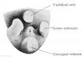
Fig. 637. Caudal end of the body of an embryo of 18 mm GL. Between the umbilicus, the coccygeal tubercle, and the right and left lower limbs is the cloacal tubercle; on its anal slope are the ostium urogenitale and the anal groove.
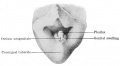
Fig. 640. The indifferent external genitalia of an embryo of 28 mm GL. The cloacal tubercle is divided into the phallus and the genital tubercles or swellings. The ostium urogenitale and anus are still close together.
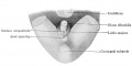
Fig. 641. Female external genitalia of an embryo of 32.5 mm GL. The phallus has become the clitoris, the glans and coronary sulcus are recognizable. The ostium urogenitale has preserved its relation to the anal opening, but no longer reaches the sulcus coronarius glandis.
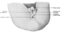
Fig. 642. Male external genitalia of an embryo of 3.5 months. The phallus has become the glans penis, the scrotal swellings are fully developed and the unpaired scrotal area is indicated. The ostium urogenitale and ostium anale have separated, the ostium urogenitale extends to the sulcus coronarius glandis.
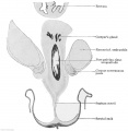
Fig. 643. Transverse section of a male embryo of 70 mm head-foot length (embryo R. Meyer 267; slide 67, row 1, section 3), below the symphysis pubis. X ca. 23. From behind forwards the following parts are cut: rectum, pars pelvina of the urogenital sinus and scrotum. From the pars pelvina Cowper's glands have developed and between A the mesenchyme of the urogenital sinus and the epithelium of the scrotum there is stretched the septum scroti.
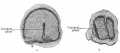
Fig. 653 a and b. Two sections through the clitoris of an embryo of 60 mm head-foot length. (Embryo R. Meyer 268; slide 66, row 2, section 1 and slide 67, row 1, section 2.) The urethral plate in section a extends from the anal surface of the clitoris only to the centre. In section 6, which passes through the tip, the clitoris is completely divided by the urethral plate.
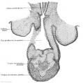
Fig. 654. Longitudinal section of the clitoris of an embryo of 60 mm head-foot length. (Embryo H. Meyer 268; slide 64, row 2, section 1.) X 50. The section cuts the sinus urogenitalis lengthwise from the crista urethralis int. to the ostium urogenitale. The lumen of the sinus is enlarged at its end to a triangular basin, on whose distal wall is the urethral plate. To the right and left are the genital swellings, the anlagen of the labia majora, which are becoming separated from the clitoris.
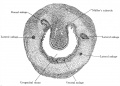
Fig. 655. Transverse section of the urogenital sinus of a male embryo of 70 mm head-foot length. (Embryo R. Meyer 267; slide 60, row 2, section 3.) X47. The section shows Mailer's tubercle projecting into the lumen of the sinus and around the sinus the anlagen of the prostatic glands.
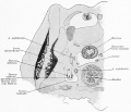
Fig. 657. Part of a transverse section through a human embryo of 60 mm. head-foot length. (Embryo R. Meyer 264; slide 44, row 2, section 2.) By the union of the mesonephric fold and testis to the anterior abdominal wall, formed by the chorda gubernaculi, both have been carried quite away from the posterior abdominal wall. Between the urogenital fold and the posterior wall a layer of loose mesenchyme has formed. Thereby the portion of the abdominal cavity which contains the urogenital fold is separated off from the rest of the cavity as the saccus vaginalis. The separation between the two is completed by a peritoneal fold formed by the a. umbilicalis.


References
Meyer, R. : Ueber die fetale Uterusschleimhaut, Zeit. f . Geburtsh. u. Gynak., vol. 38, 1898. Meyer, R. : Zur Entstehung des doppelten Uterus, Zeit. f . Geburtsh. u. Gynak., vol. 38, 1898b.
Meyer, R. : Ueber epitheliale Gebilde im Myometrium des fetalen und kindlichen Uterus einsehl. des Gartner'schen Ganges, Berlin, 1899.
Meyer, R. : Ueber sogenannte Vornierenreste und das nephrogene Zwischenblastem bei mensehlichen Embryonen und ihre eventuelle pathologische Persistenz, Charite-Annalen, vol. 33, p. 649-656, with 2 text-figs., 1909.
Meyer, R. : Zur Kenntnis des Gartner'schen Ganges besonders in der Vagina und dem Hymen des Menschen, Arch. f. mikr. Anat., vol. 73, 1909.
Meyer, R. : Zur Entwicklungsgeschichte und Anatomie des utrieulus prostaticus beim Menschen, Arch. f. mikr. Anat. u. Entw., vol. 74, 1909b.
Meyer, R. : Ueber erosio portionis uteri, Deut. patholog. Gesellsch. Erlangen, 1910.
Meyer, R.: Die Erosion und Pseudoerosion des Erwachsenen, Arch. f. Gynak. vol. 91, part. 3, p. 1-35 with 1 plate and 3 text-figs., 1910.
Meyer, R. : Die Epithelentwicklung der cervix und portio vaginalis und die pseudoerosio congenita (Kongenital. histol. Ectropium), Arch. f. Gynak., vol. 90, part 3, p. 1-20 with 1 plate, 1910.
Meyer, R. : Beitrag zur Frage nach der Genese der im Urogenitalgebiet vorkommenden Mischgeschwiilste und Teratome, Reprint from the Charite Annalen, vol. 34, p. 1-23 with 10 text-figs., 1910.
Cite this page: Hill, M.A. (2024, April 19) Embryology Embryology History - Robert Meyer. Retrieved from https://embryology.med.unsw.edu.au/embryology/index.php/Embryology_History_-_Robert_Meyer
- © Dr Mark Hill 2024, UNSW Embryology ISBN: 978 0 7334 2609 4 - UNSW CRICOS Provider Code No. 00098G