Embryology History - Orlando Charnock Bradley: Difference between revisions
(Created page with "{{Header}} ==Introduction== Orlando Charnock Bradley FRSE (1871 – 1937) was a British veterinarian and first President of the National Veterinary Medical Association. {...") |
m (→References) |
||
| (47 intermediate revisions by the same user not shown) | |||
| Line 1: | Line 1: | ||
{{Header}} | {{Header}} | ||
==Introduction== | ==Introduction== | ||
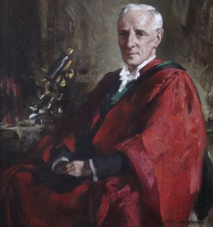
[[File:Orlando Charnock Bradley.jpg|thumb|alt=Embryology History - Orlando Charnock Bradley|link=Embryology History - Orlando Charnock Bradley|O. Charnock Bradley (1871 – 1937)]] | |||
Orlando Charnock Bradley FRSE (1871 – 1937) was a British veterinarian and first President of the National Veterinary Medical Association. | Professor Orlando Charnock Bradley FRSE (1871 – 1937) was a British veterinarian at the University of Edinburgh and first President of the National Veterinary Medical Association. | ||
He attended classes in chemistry, animal physiology and botany at the Harris Institute, Preston, before entering the New Veterinary College, Edinburgh, which was then carried on as a private enterprise by the late Professor William Williams, but was afterwards incorporated by the University of Liverpool in which it now forms the School of Veterinary Science. | |||
He was Goodsir Memorial Fellow in the years 1903-5, the title of his thesis being "The Comparative Anatomy of the Mammalian Cerebellum", and in 1905 was awarded the degree of Doctor of Science in respect of his work on the development and morphology of the mammalian hind-brain. | |||
(above text modified from obituary notice) | |||
{{History People}} | {{History People}} | ||
==Obituary Notice== | |||
Orlando Charnock Bradley was born at [https://goo.gl/maps/2YrFg5bUNWqeD8vB9 Wheelton], near Chorley, in Lancashire, on May 8, 1871. | |||
He received his early education at Wheelton, afterwards proceeding to Chorley Grammar School. Later he attended classes in chemistry, animal physiology and botany at the Harris Institute, Preston, before entering the New Veterinary College, Edinburgh, which was then carried on as a private enterprise by the late Professor William Williams, but was afterwards incorporated by the University of Liverpool in which it now forms the School of Veterinary Science. Bradley’s student career was such that his outstanding ability quickly attracted the attention of Williams, and upon his graduating M.R.C.V.S. in 1892 he was at once appointed Lecturer in Comparative Anatomy and in addition was placed in charge of the College Hospital. It cannot be said that his bent ever lay towards clinical veterinary practice, and one may venture to assume that he did not find his hospital duties particularly attractive, especially when they had to be undertaken in conjunction with his studies in comparative anatomy, in which subject, from his first contact with it, he knew he had found his true scientific sphere. It was nevertheless in the field of clinical veterinary medicine that the spirit of inquiry and the faculty of precise observation which he possessed, and in later years so markedly developed, first became evident in a published paper, for it was at this early stage that he recorded his observations on the treatment of intestinal tympany in the horse by antizymotic agents introduced directly into the colon by intestinal puncture. | |||
By reason of temperament and inclination Bradley soon became almost wholly immersed in his anatomical work, and in order (among other reasons) to extend his knowledge of comparative anatomy took up the study of medicine at the University of Edinburgh, where he graduated M.B., Ch.B., in 1900. He was Goodsir Memorial Fellow in the years 1903-5, the title of his thesis being The Comparative Anatomy of the Mammalian [[Neural - Cerebellum Development|Cerebellum]], and in 1905 was awarded the degree of Doctor of Science in respect of his work on the development and morphology of the mammalian {{hindbrain}}. Two years later there followed the conferment of his Doctorate in Medicine for his researches in the development of the mammalian {{liver}}, and in 1908 he received from the Royal College of Veterinary Surgeons the John Henry Steel Memorial Medal ‘“‘in reward of merit.” | |||
Between 1893 and 1908 Bradley’s contribution to the literature was extensive and during this period he published forty-seven original scientific communications, principally on anatomical, genetical and historical subjects. His text-books have proved of incalculable value to veterinary science. The Outlines of Veterinary Anatomy published in three parts in 1896-97 was an immature effort in which he, in after years, found no satisfaction, but his later works were wholly admirable and have become standard texts. These are: [[Embryology_History_-_Orlando_Charnock_Bradley#Topographical_Anatomy_of_the_Limbs_of_the_Horse|'''Topographical Anatomy of the Limbs of the Horse''']] (1920); [[Embryology_History_-_Orlando_Charnock_Bradley#The_Topographical_Anatomy_of_the_Thorax_and_Abdomen_of_the_Horse|'''Topographical Anatomy of the Thorax and Abdomen of the Horse''']] (1922); [[Embryology_History_-_Orlando_Charnock_Bradley#The_Topographical_Anatomy_of_the_Head_and_Neck_of_the_Horse|'''Topographical Anatomy of the Head and Neck of the Horse''']] (1923); [[Embryology_History_-_Orlando_Charnock_Bradley#Topographical_Anatomy_of_the_Dog|'''Topographical Anatomy of the Dog''']], in three editions (1919, 1927, 1935); and [[Embryology_History_-_Orlando_Charnock_Bradley#The_Structure_of_the_Fowl|'''The Structure of the Fowl''']], in two editions (1915, 1937). | |||
He founded and was editor of the excellent but short-lived Veterinary Review (1917-20), which had as its object the provision of an account of current veterinary literature of interest to the specialist and the practitioner. | |||
In his scientific writings Bradley’s diction is simple, clean-cut and explicit, but on occasion he would write, as he would often speak, with grace and charm. His Azstory of the Edinburgh Veterinary College, published on the occasion of the centenary of the College in 1923, is not only an authoritative historical record but a fascinating tale. | |||
In October 1900 he was elected to the Chair of Anatomy in the Royal (Dick) Veterinary College and to the Principalship of the College in 1911, in which year he was also appointed to the Barclay and Goodsir Lectureship in Comparative Anatomy in the University of Edinburgh. In the same year he was elected to the Council of the Royal College of Veterinary Surgeons, the governing body of the veterinary profession. | |||
This time not only marked an epoch in Bradley’s career, but the circumstances of it effected a widening of his sphere of influence and occasioned a redirection of the main lines of his activities. His research work gave place to administration not only in that which concerned: his College, but in the wider field of professional politics. His wisdom in council was early recognised by his being elected Vice-President of the Royal College in 1912, and again in 1919, before proceeding to the Presidential Chair, which he occupied from 1920 to 1922. In 1935 he received the highest honour which it is within the power of the Royal College to bestow—that of Honorary Fellowship. | |||
In 1909 Bradley in a presidential address to the Scottish Metropolitan Veterinary Medical Society outlined his conception of uniting all the many veterinary societies scattered throughout the country into one national association. At a conference held the following year the principle of amalgamation was approved, and a committee under Bradley’s chairmanship was appointed to advance the scheme. After years of labour and in face of much discouragement, approval and acceptance of the constitution and rules of that body now known as the National Veterinary Medical Association of Great Britain and Ireland were finally obtained. Bradley was first elected President in 1914 and served as President continuously until 1922. | |||
The Board of Management of the Royal (Dick) Veterinary College as early as 1907 recognised the need for the removal of the College to new and much more commodious buildings, but because of numerous difficulties and delays the actual building of the new College in Summerhall was not begun till 1913, and its memorial foundation-stone was laid two days before the outbreak of the Great War. The lean years of the war constituted a period during which his dominant qualities, quiet steadfastness to purpose and unswerving determination were most clearly evident. Not only did he find ways and means of carrying on the building of the fabric of the new College in spite of the diminution in the building funds due to the rapidly rising costs, but maintained, at least in being, the National Veterinary Association which almost from its inception he had to guide with a steady hand along the very edge of financial bankruptcy. The fine buildings in which the Edinburgh Veterinary College is now housed and the strength to which the National Veterinary Association has so rapidly attained must have been causes of much satisfaction. | |||
His interest in general scientific matters is reflected in his long association with the Royal Physical Society of Edinburgh, to the Fellowship of which he was elected in 1893, serving as Secretary 1903-11 and as President 1912-15. Between Igo1 and 1928 he made twelve contributions to the Society’s Proceedings. | |||
Bradley was elected a Fellow of the Royal Society of Edinburgh in 1903. He was a member of the Council, 1907-10 and again 1915-17, and served as Vice-President 1934-37. Two of his papers are published in the Society’s Proceedings: ‘“‘ Abdominal Viscera of Cercocebus fuliginosus and Lagothrix humboldt: (1903), and ‘‘Craniometrical Observations on the Skull of Eguus prjevalskit and other Horses” (1907). | |||
Bradley could upon occasion affect a charming geniality, but his natural manner was placid, suave, precise and even cold. His sense of humour was deep and very real, but his wit, while nimble and adroit, could be sharp. His tastes were artistic and blended curiously but smoothly with his scientific attributes. He performed with acceptance upon the violoncello and appreciated music very warmly; yet, with complete detachment, he could informedly discourse upon the mechanism of its production. He was readily touched by natural beauty, especially the beauty of flowers, and this but served to quicken his interest in the principles and practice of scientific plant-breeding. It was in the possession of so many and such different qualities and of a mind so keen, cultured, and so severally faceted that Bradley was equipped splendidly for carrying through the many important tasks which he sought and to which he set himself. The passage of time alone will permit of the proper assessment of the remarkable contribution he made to the advancement of Veterinary Science during, perhaps, the most critical phase of its development. | |||
He died on November 21, 1937. | |||
==Books== | |||
===Topographical Anatomy of the Limbs of the Horse=== | |||
'''Preface''' | |||
Were it not that the conventional preface affords a means by which an author may convey his grateful thanks to those from whom he has received advice and assistance, the present book would have been sent forth without the usual preamble. Seeing, however, that the illustrations form a highly important, helpful, and essential part of the book, it would be worse than ungrateful to neglect this opportunity to thank Mr. James T. Murray for the infinite care he has bestowed upon the drawing of the various dissections. The publishers were indeed fortunate in securing the services of one whose long experience in the illustration of works on anatomy has given him knowledge that, combined with an artistic skill it would be impertinence in me to praise, places him in the forefront of anatomical illustrators. It is greatly to be deplored that the labour difficulties of peace have not spared the workshop of the blockmakers, but have resulted in the frequent neglect of Mr. Murray's instructions respecting the reduction in size of his drawings. Some of the figures have been reduced beyond the carefully determined scale indicated on the originals, and some companion figures have not been reduced to scale. Unfortunately, these errors could have been rectified only at much expense and after long delay. | |||
To Dr. E. B. Jamieson, of the University of Edinburgh, thanks are due for help in the revision of the footnotes in which the derivation of anatomical terms is indicated. It is hoped that these brief notes will lead the student — and possibly others — to realise that terms are more than mere collections of letters. | |||
Finally, I cannot abstain from thanks to the publishers, who have been most generous in meeting all suggestions, and in permitting the free use of illustrations. | |||
0. C. B. | |||
November 1919. | |||
:'''Links:''' [https://archive.org/details/cu31924000364822/page/n8/mode/2up Internet Archive] | |||
===The Topographical Anatomy of the Thorax and Abdomen of the Horse=== | |||
'''Preface''' | |||
The plan of the present book is the same as that of its predecessor on The Topographical Anatomy of the Limbs of the Horse. Its aim is also the same, namely, to serve as a sufficiently full dissection-guide for the student, and, possibly, to help the practitioner when in doubt respecting the topography of a region. | |||
My thanks are gratefully tendered to Mr. James T. Murray for again expending great care and patience on the preparation of drawings of dissections; to my colleague, Mr. T. Grahame, M.R.C.V.S., for help when reading the proof-sheets, and at other times ; and to the publishers for their generosity in allowing a goodly number of illustrations, and for meeting my wishes in so far as the existing difficult circumstances have permitted. | |||
O. C. B. | |||
April , 1922. | |||
'''Contents''' | |||
# The Thorax | |||
# Arteries of the Thorax | |||
# The Abdomen | |||
# The Pelvic Cavity | |||
# The Female Pelvis | |||
# Arteries of the Abdomen | |||
# Nerve and Blood Supply OF THE Muscles of the Thorax and Abdomen . | |||
# Lymph Glands of the Thorax and Abdomen | |||
# The Foetal Circulation | |||
{| class="wikitable mw-collapsible mw-collapsed" | |||
! The Foetal Circulation | |||
|- | |||
| | |||
During the period when the embryo is developing in utero, the | |||
respiratory and digestive organs do not function. Some special and temporary provision is therefore necessary whereby oxygenation of the blood and a supply of nutritive material may be assured. A highly vascular placenta is formed partly by the extra-embryonic portion of the ovum, and partly by the mucous membrane of the uterus of the mother. An umbilical cord connects the placenta with the umbilicus of the embryo, and in the cord are contained the umbilical vessels through which the blood passes from the embryo to the placenta and back again. 1 | |||
1 For an account of the early development of the circulation in the embryo, one of the standard works on embryology should be consulted. | |||
Fig. 83. Foetal circulation in the foal | |||
The blood is returned from the placenta to the embryo by two vessels that, in the horse, fuse into a single umbilical vein. This follows the free margin of the falciform ligament to the liver, where it joins the portal vein. In the majority of mammals the termination of the umbilical vein is connected with the caudal vena cava by a ductus venosus, in addition to the union effected with the portal vein. In the majority of mammals, therefore, some of the blood carried by the umbilical vein traverses the liver mixed with the blood that has been gathered from the intestines, stomach, spleen and pancreas; and some is conveyed by the ductus venosus direct to the vena cava. In the horse, the absence of a ductus venosus necessitates the passage through the liver of all the blood of the umbilical vein. The origin and course of the caudal vena cava is the same in the embryo as in the adult. Its opening into the right atrium, however, is directly facing the foramen ovale, an opening in the inter-atrial septum, and it appears probable that the blood brought to the heart by the caudal vena cava passes through the foramen into the left atrium. | |||
Fig. 84. — Diagram of the foetal circulation. | |||
Fig. 85. — The foetal heart viewed from the left. The ductus arteriosus has been opened, and the arrows indicate the direction in which the blood flows. | |||
Blood is collected from the head and neck, thoracic limbs and part of the wall of the thorax of the embryo in the same manner as in the adult, and consequently is carried to the right atrium by the cranial vena cava. From the right atrium it is sent into the right ventricle, and thence into the pulmonary artery. This artery furnishes a branch to each lung as in the adult; but, the lungs being unexpanded and as yet functionless, the greater part of the blood of the pulmonary artery is short-circuited into the aorta through the ductus arteriosus. | |||
The blood of the caudal vena cava, gaining the left atrium by the foramen ovale, is there mixed with the relatively small quantity of blood brought from the lungs by the pulmonary veins. The mixed blood is injected from the atrium into the left ventricle and thence into the aorta. By the branches of the aorta it is distributed to all parts of the body, except the lungs, as it is in the adult; but from each hypo¬ gastric artery there arises an umbilical artery that leaves the body of the embryo by the umbilicus and ends in the placenta. | |||
It is apparent that the purest and most highly oxygenated blood in the embryo, as well as that richest in nutritive material, is that carried by the umbilical vein to the liver, where it is mixed with the portal venous blood. A further mixture with impure blood occurs when that of the caudal vena cava meets the pulmonary blood in the left atrium. A final contamination occurs in the aorta where the ductus arteriosus adds the blood that has been gathered by the cranial vena cava and transmitted through the right heart into the pulmonary artery. The aorta of the embryo, therefore, contains blood that is most heavily loaded with waste products and poorest in oxygen and nutritive, material. | |||
The changes that take place in the circulation at birth are as follows: | |||
With the rupture of the umbilical cord blood ceases to fiow through the umbilical arteries and vein. The former become the rounded cords (round ligaments) found in the free margin of the lateral umbilical folds of the urinary bladder of the adult, while the remains of the umbilical vein constitutes the round ligament of the liver. With the establishment of respiration the lungs are distended, and the branches of the pulmonary artery become sufficiently large to permit the passage through the lungs of all the blood ejected from the right ventricle. Blood ceases to fiow through the ductus arteriosus, which loses its lumen and becomes the ligamentum arteriosum. At birth, also, the foramen ovale closes, and the blood carried to the heart by the caudal vena cava is then compelled to enter the right atrium. In those animals in which there is a ductus venosus in the embrvo, its lumen is occluded at birth, and a ligamentum venosum is produced. | |||
|} | |||
:'''Links:''' [https://archive.org/details/b29820091/page/n8/mode/2up Internet Archive] | |||
===The Topographical Anatomy of the Head and Neck of the Horse=== | |||
'''Preface''' | |||
The plan and aim of the present instalment of an account of the topographical anatomy of the horse are the same as those according to which the volumes on the limbs and the thorax and abdomen were prepared. It is hoped that the three volumes together will furnish, in sufficient detail but without over-elaboration, a description of the form and disposition of the parts of the horse's body that will help the student, and, perchance, the practitioner. | |||
My grateful thanks are again tendered to Mr. James T. Murray for the time and care devoted to the preparation of the illustrations ; to my colleague, Mr. T. Grahame, M.R.C.V.S., for assistance in reading the proof-sheets ; and to the publishers. | |||
0. C. B. | |||
September, 1923. | |||
:'''Links:''' [https://archive.org/details/topographicalanahn00brad/page/n1/mode/2up Internet Archive] | |||
===The Structure of the Fowl=== | |||
'''Preface''' | |||
Stated briefly, the primary object in view in the preparation of the following pages has been the production of a concise and not too elaborate account of naked-eye and microscopic structure which, it is hoped, may be of use to those who have to deal with the fowl in health and disease. It is a truism that to the student of avian and comparative pathology a knowledge of normal structure is essential to the appreciation of changes resulting from disease. And it is admitted that the student of scientific agriculture should know something of the build of the various animal machines with which he is concerned. | |||
It is also hoped that the student of zoology and comparative anatomy may find herein a useful introduction to avian anatomy and embryology. | |||
The main difficulty experienced by the writer has been the determination of what to include and what to leave out, having regard to the fact that a student's manual was planned and not a specialist's book of reference. In deciding the scope of the book, much valuable help has been received in the form of suggestions made by friends of considerable teaching experience. To these and to Dr. John D. Comrie, the General Editor of the Series, I tender my thanks, and desire also to express my appreciation of the unfailing courtesy of the publishers. | |||
O. CHARNOCK BRADLEY. | |||
Edinburgh, 1915. | |||
:'''Links:''' [https://archive.org/details/structureoffowl00brad/page/n9/mode/2up Internet Archive] | |||
===Topographical Anatomy of the Dog=== | |||
'''Preface to the Second Edition''' | |||
Since the first edition of this book appeared under the title of “ Notes on the Dissection of the Dog,” notable advance has been made in canine surgery. In his work in general, and more particularly in the methods and the character of his operations on the smaller domestic animals, the veterinary surgeon of to-day is following as closely as may be the specialist in human surgery ; and there is no obvious reason why human and canine surgery should not be even more nearly alike. It may be reasonably expected that the canine surgeon should, if necessary, be able to perform operations on the dog similar to any that the human surgeon performs on man. But clearly a fundamental and working knowledge of anatomy must precede*the practice of surgery, and it is hoped that the present publication may be of service in this direction. | |||
Though prepared, in the first instance, for the guidance of those who propose to practise canine surgery, it is possible that the following pages may prove useful to the student of comparative anatomy also. Indeed, it is known that the first edition did serve such a purpose. | |||
Experience gained by the use of the previous edition as a class-book has shown the desirability of certain alterations in arrangement that have been made in the present issue. The whole text, naturally, has been subjected to revision, and some parts have been entirely rewritten. A number of new illustrations have been added. These have been drawn by Mr James T. Murray, an artist of outstanding eminence as an illustrator of works on anatomy, for whose great care and unbounded patience thanks are here tendered. | |||
Acknowledgment of indebtedness must be made for valuable hints received from time to time from my colleagues; and thanks are due also to those who, having used the first edition, have pointed out inconveniences of arrangement and infelicities and inaccuracies of statement. To my colleague, Mr T. Graharne, F.R.C.V.S., especial thanks must be given for invaluable suggestions and help at all stages in the preparation of the book for the press. | |||
O. CHARNOCK BRADLEY. | |||
'''Review''' - {{J Anat.}} 1936 Jan; 70(Pt 2): 322. | |||
(3rd ed.) By O. Charnock Bradley, M.D., D.Sc., F.R.C.V.S. (Edinburgh: Oliver and Boyd, Ltd.) 1935. Pp.284,figs.91. Price 25s. net. | |||
This distinguished practical manual of the dissection of the dog is now in its third edition. The text is lucid, adequate and direct. The instructions for dissection are simple, clear and convenient. The illustrations, which number 91, are excellent, and many are in colour. Anyone - veterinary surgeon, laboratory worker, comparative anatomist - who needs to know or orientate himself in the anatomy of the dog, has in Professor Bradley's book the most complete and handiest guide he could wish for. | |||
:'''Links:''' [https://archive.org/details/b29820108/mode/2up Internet Archive] | |||
===History of the Edinburgh Veterinary College=== | |||
'''Preface''' | |||
There are good reasons why a history of the Edinburgh veterinary school should be written, and even better ones why it should be written at the present time. The life of the Founder is inspiring, for the history of his school is very different from that of the majority of institutions devoted to education. No wealthy man endowed it: no public subscription-list brought it into being: no appeal was made to or support given by the State : a famous University could not help. A poor man, the son of poor parents, started out on a venture supported by nothing more than a promise of £ 50 . He cut his coat according to his cloth, and continued so to cut it—weaving the cloth himself. And there is something almost romantic in the story of a brother and sister, living in and for the Clyde Street school, tending and watching over it continually, proudly noting its growth, doubtless exulting a little as they saw it go from strength to strength, and, dying, making it their sole heir. The picture is enriched by the figure of William Worthington, that loyal and devoted servant who so well deserves more generous treatment than he has received in this history. The desire to do him justice was there, but material facts were wanting. | |||
Further, it is fitting that as complete a story of the school as is possible should be compiled at the end of the first century of its existence. The history of the College up to the time of the Founder’s death was written in 1869 by R. O. Pringle, as an Introduction to Occasional Payers , but the later chapters can only be read in the pages of Minute Books. | |||
I dare not hope that every detail in the following pages is free from error, but it can be claimed that no pains have been spared to verify statements so far as this has been possible. | |||
It was my original intention to include a chapter in which the names and achievements of notable alumni should be recorded. The task, however, was hardly begun before difficulties made themselves manifest. With the truly notable dead there was no trouble: everyone would have agreed that their inclusion was justified. But where was the line to be drawn? Who was to say that this or that departed alumnus had or had not attained real notability ? And if differences of opinion were inevitable in the case of those whose career is closed, even more certain is it that there would have been disagreement respecting those who have yet, perchance, to add many leaves to their bays. The attempt, therefore, was abandoned. If the future holds the making of any list of graduates — and it should —the list must be a complete one. It must contain names that are familiar far beyond the confines of the veterinary profession, and names known only to intimates. This means that it will have to be compiled and published entirely con amove. A gladly willing compiler can be found; but a publisher ----- | |||
Grateful acknowledgments are here made to all those — and they are many — who have helped in the preparation of this short story. Very special thanks are due to my colleague, Professor J. Russell Greig, who has given time and care liberally to the elucidation of doubtful points. To him I also owe thanks for many fruitful suggestions and hints, and help in directions too numerous to specify. Thanks are also due to Mr A. Grierson, Town Clerk of the City of Edinburgh, and to his assistant Mr J. Jarvis, for facilities and help in connection with the examination of the Minute Books containing the history of the College from 1866 to 1906; to Mr Fred Bullock, Librarian of the Royal College of Veterinary Surgeons, for information relating to Dick’s connection with the R.C.V.S., and for the dates of the foundation of the European veterinary schools; to Mr John Stirton, Secretary of the Highland and Agricultural Society, for help in verifying the earlier history of the College; to Mr J. T. Murray for the care with which he has made the drawing of the new College buildings; to the Council of the Royal College of Physicians of Edinburgh for permission to copy the portrait of Dr John Barclay ; to Principal Hewlett, of the Bombay Veterinary College, to Sir Stewart Stockman, and to Mr J. Clarkson, for portraits of former Principals of the College; to Mr R. P. Phillimore for the loan of a block of his etching “White Horse Close”; to Messrs A. & C. Black for permission to copy the engraving of “Paul’s Work”; and to the staffs of the North British Agriculturist , the Scotsman , and the Register House for help in consulting files and records. | |||
O. Charnock Bradley. | |||
:'''Links:''' [https://archive.org/details/b29827176/page/n9/mode/2up Internet Archive] | [https://www.ed.ac.uk/vet The Royal (Dick) School of Veterinary Studies] | |||
==References== | ==References== | ||
{{Ref-Bradley1903a}} | |||
{{Ref-Bradley1903b}} | |||
{{Ref-Bradley1904a}} | |||
{{Ref-Bradley1904b}} | |||
{{Ref-Bradley1905}} | |||
{{Ref-Bradley1906}} | |||
{{Ref-Bradley1908}} | |||
{{#pmid:17232788}} | |||
{{#pmid:17232671}} | |||
{{#pmid:17232657}} | |||
{{#pmid:17232628}} | |||
{{#pmid:17232617}} | |||
{{#pmid:17232554}} | |||
{{#pmid:17104198}} | |||
Search PubMed: [https://www.ncbi.nlm.nih.gov/pubmed/?term=Bradley%20OC%5BAuthor%5D&cauthor=true&cauthor_uid=17232548 Orlando Charnock Bradley] | Search PubMed: [https://www.ncbi.nlm.nih.gov/pubmed/?term=Bradley%20OC%5BAuthor%5D&cauthor=true&cauthor_uid=17232548 Orlando Charnock Bradley] | ||
Search Internet Archive: [https://archive.org/search.php?query=creator%3A%22Bradley%2C+O.+Charnock Orlando Charnock Bradley] | |||
{{Footer}} | {{Footer}} | ||
[[Category:People]][[Category:United Kingdom]] | [[Category:People]][[Category:United Kingdom]] | ||
[[Category:Historic Embryology]][[Category:1900's]] | [[Category:Historic Embryology]][[Category:1900's]] | ||
[[Category:Neural]] | [[Category:Neural]][[Category:Draft]] | ||
Latest revision as of 11:10, 14 May 2020
| Embryology - 20 Apr 2024 |
|---|
| Google Translate - select your language from the list shown below (this will open a new external page) |
|
العربية | català | 中文 | 中國傳統的 | français | Deutsche | עִברִית | हिंदी | bahasa Indonesia | italiano | 日本語 | 한국어 | မြန်မာ | Pilipino | Polskie | português | ਪੰਜਾਬੀ ਦੇ | Română | русский | Español | Swahili | Svensk | ไทย | Türkçe | اردو | ייִדיש | Tiếng Việt These external translations are automated and may not be accurate. (More? About Translations) |
Introduction
Professor Orlando Charnock Bradley FRSE (1871 – 1937) was a British veterinarian at the University of Edinburgh and first President of the National Veterinary Medical Association.
He attended classes in chemistry, animal physiology and botany at the Harris Institute, Preston, before entering the New Veterinary College, Edinburgh, which was then carried on as a private enterprise by the late Professor William Williams, but was afterwards incorporated by the University of Liverpool in which it now forms the School of Veterinary Science.
He was Goodsir Memorial Fellow in the years 1903-5, the title of his thesis being "The Comparative Anatomy of the Mammalian Cerebellum", and in 1905 was awarded the degree of Doctor of Science in respect of his work on the development and morphology of the mammalian hind-brain.
(above text modified from obituary notice)
| Embryologists: William Hunter | Wilhelm Roux | Caspar Wolff | Wilhelm His | Oscar Hertwig | Julius Kollmann | Hans Spemann | Francis Balfour | Charles Minot | Ambrosius Hubrecht | Charles Bardeen | Franz Keibel | Franklin Mall | Florence Sabin | George Streeter | George Corner | James Hill | Jan Florian | Thomas Bryce | Thomas Morgan | Ernest Frazer | Francisco Orts-Llorca | José Doménech Mateu | Frederic Lewis | Arthur Meyer | Robert Meyer | Erich Blechschmidt | Klaus Hinrichsen | Hideo Nishimura | Arthur Hertig | John Rock | Viktor Hamburger | Mary Lyon | Nicole Le Douarin | Robert Winston | Fabiola Müller | Ronan O'Rahilly | Robert Edwards | John Gurdon | Shinya Yamanaka | Embryology History | Category:People | ||
|
Obituary Notice
Orlando Charnock Bradley was born at Wheelton, near Chorley, in Lancashire, on May 8, 1871.
He received his early education at Wheelton, afterwards proceeding to Chorley Grammar School. Later he attended classes in chemistry, animal physiology and botany at the Harris Institute, Preston, before entering the New Veterinary College, Edinburgh, which was then carried on as a private enterprise by the late Professor William Williams, but was afterwards incorporated by the University of Liverpool in which it now forms the School of Veterinary Science. Bradley’s student career was such that his outstanding ability quickly attracted the attention of Williams, and upon his graduating M.R.C.V.S. in 1892 he was at once appointed Lecturer in Comparative Anatomy and in addition was placed in charge of the College Hospital. It cannot be said that his bent ever lay towards clinical veterinary practice, and one may venture to assume that he did not find his hospital duties particularly attractive, especially when they had to be undertaken in conjunction with his studies in comparative anatomy, in which subject, from his first contact with it, he knew he had found his true scientific sphere. It was nevertheless in the field of clinical veterinary medicine that the spirit of inquiry and the faculty of precise observation which he possessed, and in later years so markedly developed, first became evident in a published paper, for it was at this early stage that he recorded his observations on the treatment of intestinal tympany in the horse by antizymotic agents introduced directly into the colon by intestinal puncture.
By reason of temperament and inclination Bradley soon became almost wholly immersed in his anatomical work, and in order (among other reasons) to extend his knowledge of comparative anatomy took up the study of medicine at the University of Edinburgh, where he graduated M.B., Ch.B., in 1900. He was Goodsir Memorial Fellow in the years 1903-5, the title of his thesis being The Comparative Anatomy of the Mammalian Cerebellum, and in 1905 was awarded the degree of Doctor of Science in respect of his work on the development and morphology of the mammalian hindbrain. Two years later there followed the conferment of his Doctorate in Medicine for his researches in the development of the mammalian liver, and in 1908 he received from the Royal College of Veterinary Surgeons the John Henry Steel Memorial Medal ‘“‘in reward of merit.”
Between 1893 and 1908 Bradley’s contribution to the literature was extensive and during this period he published forty-seven original scientific communications, principally on anatomical, genetical and historical subjects. His text-books have proved of incalculable value to veterinary science. The Outlines of Veterinary Anatomy published in three parts in 1896-97 was an immature effort in which he, in after years, found no satisfaction, but his later works were wholly admirable and have become standard texts. These are: Topographical Anatomy of the Limbs of the Horse (1920); Topographical Anatomy of the Thorax and Abdomen of the Horse (1922); Topographical Anatomy of the Head and Neck of the Horse (1923); Topographical Anatomy of the Dog, in three editions (1919, 1927, 1935); and The Structure of the Fowl, in two editions (1915, 1937).
He founded and was editor of the excellent but short-lived Veterinary Review (1917-20), which had as its object the provision of an account of current veterinary literature of interest to the specialist and the practitioner.
In his scientific writings Bradley’s diction is simple, clean-cut and explicit, but on occasion he would write, as he would often speak, with grace and charm. His Azstory of the Edinburgh Veterinary College, published on the occasion of the centenary of the College in 1923, is not only an authoritative historical record but a fascinating tale.
In October 1900 he was elected to the Chair of Anatomy in the Royal (Dick) Veterinary College and to the Principalship of the College in 1911, in which year he was also appointed to the Barclay and Goodsir Lectureship in Comparative Anatomy in the University of Edinburgh. In the same year he was elected to the Council of the Royal College of Veterinary Surgeons, the governing body of the veterinary profession.
This time not only marked an epoch in Bradley’s career, but the circumstances of it effected a widening of his sphere of influence and occasioned a redirection of the main lines of his activities. His research work gave place to administration not only in that which concerned: his College, but in the wider field of professional politics. His wisdom in council was early recognised by his being elected Vice-President of the Royal College in 1912, and again in 1919, before proceeding to the Presidential Chair, which he occupied from 1920 to 1922. In 1935 he received the highest honour which it is within the power of the Royal College to bestow—that of Honorary Fellowship.
In 1909 Bradley in a presidential address to the Scottish Metropolitan Veterinary Medical Society outlined his conception of uniting all the many veterinary societies scattered throughout the country into one national association. At a conference held the following year the principle of amalgamation was approved, and a committee under Bradley’s chairmanship was appointed to advance the scheme. After years of labour and in face of much discouragement, approval and acceptance of the constitution and rules of that body now known as the National Veterinary Medical Association of Great Britain and Ireland were finally obtained. Bradley was first elected President in 1914 and served as President continuously until 1922.
The Board of Management of the Royal (Dick) Veterinary College as early as 1907 recognised the need for the removal of the College to new and much more commodious buildings, but because of numerous difficulties and delays the actual building of the new College in Summerhall was not begun till 1913, and its memorial foundation-stone was laid two days before the outbreak of the Great War. The lean years of the war constituted a period during which his dominant qualities, quiet steadfastness to purpose and unswerving determination were most clearly evident. Not only did he find ways and means of carrying on the building of the fabric of the new College in spite of the diminution in the building funds due to the rapidly rising costs, but maintained, at least in being, the National Veterinary Association which almost from its inception he had to guide with a steady hand along the very edge of financial bankruptcy. The fine buildings in which the Edinburgh Veterinary College is now housed and the strength to which the National Veterinary Association has so rapidly attained must have been causes of much satisfaction.
His interest in general scientific matters is reflected in his long association with the Royal Physical Society of Edinburgh, to the Fellowship of which he was elected in 1893, serving as Secretary 1903-11 and as President 1912-15. Between Igo1 and 1928 he made twelve contributions to the Society’s Proceedings.
Bradley was elected a Fellow of the Royal Society of Edinburgh in 1903. He was a member of the Council, 1907-10 and again 1915-17, and served as Vice-President 1934-37. Two of his papers are published in the Society’s Proceedings: ‘“‘ Abdominal Viscera of Cercocebus fuliginosus and Lagothrix humboldt: (1903), and ‘‘Craniometrical Observations on the Skull of Eguus prjevalskit and other Horses” (1907).
Bradley could upon occasion affect a charming geniality, but his natural manner was placid, suave, precise and even cold. His sense of humour was deep and very real, but his wit, while nimble and adroit, could be sharp. His tastes were artistic and blended curiously but smoothly with his scientific attributes. He performed with acceptance upon the violoncello and appreciated music very warmly; yet, with complete detachment, he could informedly discourse upon the mechanism of its production. He was readily touched by natural beauty, especially the beauty of flowers, and this but served to quicken his interest in the principles and practice of scientific plant-breeding. It was in the possession of so many and such different qualities and of a mind so keen, cultured, and so severally faceted that Bradley was equipped splendidly for carrying through the many important tasks which he sought and to which he set himself. The passage of time alone will permit of the proper assessment of the remarkable contribution he made to the advancement of Veterinary Science during, perhaps, the most critical phase of its development.
He died on November 21, 1937.
Books
Topographical Anatomy of the Limbs of the Horse
Preface
Were it not that the conventional preface affords a means by which an author may convey his grateful thanks to those from whom he has received advice and assistance, the present book would have been sent forth without the usual preamble. Seeing, however, that the illustrations form a highly important, helpful, and essential part of the book, it would be worse than ungrateful to neglect this opportunity to thank Mr. James T. Murray for the infinite care he has bestowed upon the drawing of the various dissections. The publishers were indeed fortunate in securing the services of one whose long experience in the illustration of works on anatomy has given him knowledge that, combined with an artistic skill it would be impertinence in me to praise, places him in the forefront of anatomical illustrators. It is greatly to be deplored that the labour difficulties of peace have not spared the workshop of the blockmakers, but have resulted in the frequent neglect of Mr. Murray's instructions respecting the reduction in size of his drawings. Some of the figures have been reduced beyond the carefully determined scale indicated on the originals, and some companion figures have not been reduced to scale. Unfortunately, these errors could have been rectified only at much expense and after long delay.
To Dr. E. B. Jamieson, of the University of Edinburgh, thanks are due for help in the revision of the footnotes in which the derivation of anatomical terms is indicated. It is hoped that these brief notes will lead the student — and possibly others — to realise that terms are more than mere collections of letters.
Finally, I cannot abstain from thanks to the publishers, who have been most generous in meeting all suggestions, and in permitting the free use of illustrations.
0. C. B.
November 1919.
- Links: Internet Archive
The Topographical Anatomy of the Thorax and Abdomen of the Horse
Preface
The plan of the present book is the same as that of its predecessor on The Topographical Anatomy of the Limbs of the Horse. Its aim is also the same, namely, to serve as a sufficiently full dissection-guide for the student, and, possibly, to help the practitioner when in doubt respecting the topography of a region.
My thanks are gratefully tendered to Mr. James T. Murray for again expending great care and patience on the preparation of drawings of dissections; to my colleague, Mr. T. Grahame, M.R.C.V.S., for help when reading the proof-sheets, and at other times ; and to the publishers for their generosity in allowing a goodly number of illustrations, and for meeting my wishes in so far as the existing difficult circumstances have permitted.
O. C. B.
April , 1922.
Contents
- The Thorax
- Arteries of the Thorax
- The Abdomen
- The Pelvic Cavity
- The Female Pelvis
- Arteries of the Abdomen
- Nerve and Blood Supply OF THE Muscles of the Thorax and Abdomen .
- Lymph Glands of the Thorax and Abdomen
- The Foetal Circulation
| The Foetal Circulation |
|---|
|
During the period when the embryo is developing in utero, the respiratory and digestive organs do not function. Some special and temporary provision is therefore necessary whereby oxygenation of the blood and a supply of nutritive material may be assured. A highly vascular placenta is formed partly by the extra-embryonic portion of the ovum, and partly by the mucous membrane of the uterus of the mother. An umbilical cord connects the placenta with the umbilicus of the embryo, and in the cord are contained the umbilical vessels through which the blood passes from the embryo to the placenta and back again. 1 1 For an account of the early development of the circulation in the embryo, one of the standard works on embryology should be consulted. Fig. 83. Foetal circulation in the foal The blood is returned from the placenta to the embryo by two vessels that, in the horse, fuse into a single umbilical vein. This follows the free margin of the falciform ligament to the liver, where it joins the portal vein. In the majority of mammals the termination of the umbilical vein is connected with the caudal vena cava by a ductus venosus, in addition to the union effected with the portal vein. In the majority of mammals, therefore, some of the blood carried by the umbilical vein traverses the liver mixed with the blood that has been gathered from the intestines, stomach, spleen and pancreas; and some is conveyed by the ductus venosus direct to the vena cava. In the horse, the absence of a ductus venosus necessitates the passage through the liver of all the blood of the umbilical vein. The origin and course of the caudal vena cava is the same in the embryo as in the adult. Its opening into the right atrium, however, is directly facing the foramen ovale, an opening in the inter-atrial septum, and it appears probable that the blood brought to the heart by the caudal vena cava passes through the foramen into the left atrium.
The blood of the caudal vena cava, gaining the left atrium by the foramen ovale, is there mixed with the relatively small quantity of blood brought from the lungs by the pulmonary veins. The mixed blood is injected from the atrium into the left ventricle and thence into the aorta. By the branches of the aorta it is distributed to all parts of the body, except the lungs, as it is in the adult; but from each hypo¬ gastric artery there arises an umbilical artery that leaves the body of the embryo by the umbilicus and ends in the placenta. It is apparent that the purest and most highly oxygenated blood in the embryo, as well as that richest in nutritive material, is that carried by the umbilical vein to the liver, where it is mixed with the portal venous blood. A further mixture with impure blood occurs when that of the caudal vena cava meets the pulmonary blood in the left atrium. A final contamination occurs in the aorta where the ductus arteriosus adds the blood that has been gathered by the cranial vena cava and transmitted through the right heart into the pulmonary artery. The aorta of the embryo, therefore, contains blood that is most heavily loaded with waste products and poorest in oxygen and nutritive, material. The changes that take place in the circulation at birth are as follows: With the rupture of the umbilical cord blood ceases to fiow through the umbilical arteries and vein. The former become the rounded cords (round ligaments) found in the free margin of the lateral umbilical folds of the urinary bladder of the adult, while the remains of the umbilical vein constitutes the round ligament of the liver. With the establishment of respiration the lungs are distended, and the branches of the pulmonary artery become sufficiently large to permit the passage through the lungs of all the blood ejected from the right ventricle. Blood ceases to fiow through the ductus arteriosus, which loses its lumen and becomes the ligamentum arteriosum. At birth, also, the foramen ovale closes, and the blood carried to the heart by the caudal vena cava is then compelled to enter the right atrium. In those animals in which there is a ductus venosus in the embrvo, its lumen is occluded at birth, and a ligamentum venosum is produced. |
- Links: Internet Archive
The Topographical Anatomy of the Head and Neck of the Horse
Preface
The plan and aim of the present instalment of an account of the topographical anatomy of the horse are the same as those according to which the volumes on the limbs and the thorax and abdomen were prepared. It is hoped that the three volumes together will furnish, in sufficient detail but without over-elaboration, a description of the form and disposition of the parts of the horse's body that will help the student, and, perchance, the practitioner.
My grateful thanks are again tendered to Mr. James T. Murray for the time and care devoted to the preparation of the illustrations ; to my colleague, Mr. T. Grahame, M.R.C.V.S., for assistance in reading the proof-sheets ; and to the publishers.
0. C. B.
September, 1923.
- Links: Internet Archive
The Structure of the Fowl
Preface
Stated briefly, the primary object in view in the preparation of the following pages has been the production of a concise and not too elaborate account of naked-eye and microscopic structure which, it is hoped, may be of use to those who have to deal with the fowl in health and disease. It is a truism that to the student of avian and comparative pathology a knowledge of normal structure is essential to the appreciation of changes resulting from disease. And it is admitted that the student of scientific agriculture should know something of the build of the various animal machines with which he is concerned.
It is also hoped that the student of zoology and comparative anatomy may find herein a useful introduction to avian anatomy and embryology.
The main difficulty experienced by the writer has been the determination of what to include and what to leave out, having regard to the fact that a student's manual was planned and not a specialist's book of reference. In deciding the scope of the book, much valuable help has been received in the form of suggestions made by friends of considerable teaching experience. To these and to Dr. John D. Comrie, the General Editor of the Series, I tender my thanks, and desire also to express my appreciation of the unfailing courtesy of the publishers.
O. CHARNOCK BRADLEY.
Edinburgh, 1915.
- Links: Internet Archive
Topographical Anatomy of the Dog
Preface to the Second Edition
Since the first edition of this book appeared under the title of “ Notes on the Dissection of the Dog,” notable advance has been made in canine surgery. In his work in general, and more particularly in the methods and the character of his operations on the smaller domestic animals, the veterinary surgeon of to-day is following as closely as may be the specialist in human surgery ; and there is no obvious reason why human and canine surgery should not be even more nearly alike. It may be reasonably expected that the canine surgeon should, if necessary, be able to perform operations on the dog similar to any that the human surgeon performs on man. But clearly a fundamental and working knowledge of anatomy must precede*the practice of surgery, and it is hoped that the present publication may be of service in this direction.
Though prepared, in the first instance, for the guidance of those who propose to practise canine surgery, it is possible that the following pages may prove useful to the student of comparative anatomy also. Indeed, it is known that the first edition did serve such a purpose.
Experience gained by the use of the previous edition as a class-book has shown the desirability of certain alterations in arrangement that have been made in the present issue. The whole text, naturally, has been subjected to revision, and some parts have been entirely rewritten. A number of new illustrations have been added. These have been drawn by Mr James T. Murray, an artist of outstanding eminence as an illustrator of works on anatomy, for whose great care and unbounded patience thanks are here tendered.
Acknowledgment of indebtedness must be made for valuable hints received from time to time from my colleagues; and thanks are due also to those who, having used the first edition, have pointed out inconveniences of arrangement and infelicities and inaccuracies of statement. To my colleague, Mr T. Graharne, F.R.C.V.S., especial thanks must be given for invaluable suggestions and help at all stages in the preparation of the book for the press.
O. CHARNOCK BRADLEY.
Review - J Anat. 1936 Jan; 70(Pt 2): 322.
(3rd ed.) By O. Charnock Bradley, M.D., D.Sc., F.R.C.V.S. (Edinburgh: Oliver and Boyd, Ltd.) 1935. Pp.284,figs.91. Price 25s. net. This distinguished practical manual of the dissection of the dog is now in its third edition. The text is lucid, adequate and direct. The instructions for dissection are simple, clear and convenient. The illustrations, which number 91, are excellent, and many are in colour. Anyone - veterinary surgeon, laboratory worker, comparative anatomist - who needs to know or orientate himself in the anatomy of the dog, has in Professor Bradley's book the most complete and handiest guide he could wish for.
- Links: Internet Archive
History of the Edinburgh Veterinary College
Preface
There are good reasons why a history of the Edinburgh veterinary school should be written, and even better ones why it should be written at the present time. The life of the Founder is inspiring, for the history of his school is very different from that of the majority of institutions devoted to education. No wealthy man endowed it: no public subscription-list brought it into being: no appeal was made to or support given by the State : a famous University could not help. A poor man, the son of poor parents, started out on a venture supported by nothing more than a promise of £ 50 . He cut his coat according to his cloth, and continued so to cut it—weaving the cloth himself. And there is something almost romantic in the story of a brother and sister, living in and for the Clyde Street school, tending and watching over it continually, proudly noting its growth, doubtless exulting a little as they saw it go from strength to strength, and, dying, making it their sole heir. The picture is enriched by the figure of William Worthington, that loyal and devoted servant who so well deserves more generous treatment than he has received in this history. The desire to do him justice was there, but material facts were wanting.
Further, it is fitting that as complete a story of the school as is possible should be compiled at the end of the first century of its existence. The history of the College up to the time of the Founder’s death was written in 1869 by R. O. Pringle, as an Introduction to Occasional Payers , but the later chapters can only be read in the pages of Minute Books.
I dare not hope that every detail in the following pages is free from error, but it can be claimed that no pains have been spared to verify statements so far as this has been possible.
It was my original intention to include a chapter in which the names and achievements of notable alumni should be recorded. The task, however, was hardly begun before difficulties made themselves manifest. With the truly notable dead there was no trouble: everyone would have agreed that their inclusion was justified. But where was the line to be drawn? Who was to say that this or that departed alumnus had or had not attained real notability ? And if differences of opinion were inevitable in the case of those whose career is closed, even more certain is it that there would have been disagreement respecting those who have yet, perchance, to add many leaves to their bays. The attempt, therefore, was abandoned. If the future holds the making of any list of graduates — and it should —the list must be a complete one. It must contain names that are familiar far beyond the confines of the veterinary profession, and names known only to intimates. This means that it will have to be compiled and published entirely con amove. A gladly willing compiler can be found; but a publisher -----
Grateful acknowledgments are here made to all those — and they are many — who have helped in the preparation of this short story. Very special thanks are due to my colleague, Professor J. Russell Greig, who has given time and care liberally to the elucidation of doubtful points. To him I also owe thanks for many fruitful suggestions and hints, and help in directions too numerous to specify. Thanks are also due to Mr A. Grierson, Town Clerk of the City of Edinburgh, and to his assistant Mr J. Jarvis, for facilities and help in connection with the examination of the Minute Books containing the history of the College from 1866 to 1906; to Mr Fred Bullock, Librarian of the Royal College of Veterinary Surgeons, for information relating to Dick’s connection with the R.C.V.S., and for the dates of the foundation of the European veterinary schools; to Mr John Stirton, Secretary of the Highland and Agricultural Society, for help in verifying the earlier history of the College; to Mr J. T. Murray for the care with which he has made the drawing of the new College buildings; to the Council of the Royal College of Physicians of Edinburgh for permission to copy the portrait of Dr John Barclay ; to Principal Hewlett, of the Bombay Veterinary College, to Sir Stewart Stockman, and to Mr J. Clarkson, for portraits of former Principals of the College; to Mr R. P. Phillimore for the loan of a block of his etching “White Horse Close”; to Messrs A. & C. Black for permission to copy the engraving of “Paul’s Work”; and to the staffs of the North British Agriculturist , the Scotsman , and the Register House for help in consulting files and records.
O. Charnock Bradley.
References
Bradley OC. On the Development and Homology of the Mammalian Cerebellar Fissures: Part I. (1903) J Anat. Physiol. 37: 112-30. PMID 17232548
Bradley OC. Development and Homology of the Mammalian Cerebellar Fissures: Part II. (1903) J Anat Physiol 37: 221-240.13. PMID 17232554
Bradley OC. The mammalian cerebellum: its lobes and fissures. (1904) J Anat Physiol. 38(4): 448-475. PMID17232617
Bradley OC. The mammalian cerebellum: its lobes and fissures. (1904) J Anat Physiol. 39(1): 99–117. PMID 17232628
Bradley OC. On the Development of the Hind-Brain of the Pig: Part I. (1905) 40, 1-14.13. PMID 17232657
Bradley OC. On the Development of the Hind-Brain of the Pig: Part II. (1905) 40, 133-151.6 PMID 17232671
Bradley OC. A contribution to the morphology and development of the mammalian liver. (1908) J Anat. 43: 1-42. PMID 17232788
Bradley OC. (1908). A Contribution to the Morphology and Development of the Mammalian Liver. J Anat Physiol , 43, 1-42. PMID: 17232788
Bradley OC. (1906). On the Development of the Hind-Brain of the Pig: Part II. J Anat Physiol , 40, 133-151.6. PMID: 17232671
Bradley OC. (1905). On the Development of the Hind-Brain of the Pig: Part I. J Anat Physiol , 40, 1-14.13. PMID: 17232657
Bradley OC. (1904). The Mammalian Cerebellum: Its Lobes and Fissures. J Anat Physiol , 39, 99-117. PMID: 17232628
Bradley OC. (1904). The Mammalian Cerebellum: Its Lobes and Fissures. J Anat Physiol , 38, 448-75. PMID: 17232617
Bradley OC. (1903). Development and Homology of the Mammalian Cerebellar Fissures: Part II. J Anat Physiol , 37, 221-240.13. PMID: 17232554
Bradley OC. (1928). Notes on the Histology of the Oviduct of the Domestic Hen. J. Anat. , 62, 339-45. PMID: 17104198
Search PubMed: Orlando Charnock Bradley
Search Internet Archive: Orlando Charnock Bradley
Cite this page: Hill, M.A. (2024, April 20) Embryology Embryology History - Orlando Charnock Bradley. Retrieved from https://embryology.med.unsw.edu.au/embryology/index.php/Embryology_History_-_Orlando_Charnock_Bradley
- © Dr Mark Hill 2024, UNSW Embryology ISBN: 978 0 7334 2609 4 - UNSW CRICOS Provider Code No. 00098G