Embryo Serial Sections
| Embryology - 25 Apr 2024 |
|---|
| Google Translate - select your language from the list shown below (this will open a new external page) |
|
العربية | català | 中文 | 中國傳統的 | français | Deutsche | עִברִית | हिंदी | bahasa Indonesia | italiano | 日本語 | 한국어 | မြန်မာ | Pilipino | Polskie | português | ਪੰਜਾਬੀ ਦੇ | Română | русский | Español | Swahili | Svensk | ไทย | Türkçe | اردو | ייִדיש | Tiếng Việt These external translations are automated and may not be accurate. (More? About Translations) |
Introduction
Historically, an important way of teaching embryology is by direct observation of histological sections at different stages of development. Even today the best way to observe anatomical development and relationships is by direct observation in serial sections through the embryo and fetus. The sections below are from the original teaching set prepared at UNSW that were digitized and put online in 1996 as a teaching resource.
The Stage 13 and 22 images are available in at least two formats, an unlabeled set (image 1 - 49) and a labeled set (image 50 - 98) in a rostro-caudal sequence (head to tail). Stage 22 embryo also has a set of selected images showing more detail of some organ and tissue development. In addition, there are now 3D animations based upon reconstruction of the embryos from these serial sections.
2011 - Selected slides are currently being rescanned at high resolution to show specific developmental details.
| Author Comments |
|---|
 Start here by looking through the early (week 4) embryo (stage 13) labeled images. It is not so important to identify every single feature, observe what structures are present and where they are in the middle of the embryonic period (week 4). Then if you like, look through the unlabeled images. What can you now recognise? Start here by looking through the early (week 4) embryo (stage 13) labeled images. It is not so important to identify every single feature, observe what structures are present and where they are in the middle of the embryonic period (week 4). Then if you like, look through the unlabeled images. What can you now recognise?
Next look through the late (week 8) embryo (stage 22) labeled images as you did before. Then look through the same embryo stage selected labeled images. These show organ and tissue development of specific structures by the end of the embryonic period. What has changed? Finally put the systems together by watching the embryo animations based upon these sections. How different are the embryonic systems to the adult anatomy? More about these original serial images and associated notes. |
- Links: stage 13 sections | Carnegie stage 13 | Movies - stage 13 | stage 22 sections stage 22 selected sections | Carnegie stage 22 | Movies - stage 22
Carnegie Stage 13
About Stage 13 Embryo Sections - This image is from a serial section of a 6mm CRL pig embryo with some features of the Stage 14 embryo. This embryo is approximately equal to the day 42 human embryo. Use these serial images to identify internal features and relationships that exist within the embryo at this stage. Then compare these images with the later features of the Carnegie stage 22 human embryo.
| Stage 13 Serial unlabeled images | Embryo Stage 13 Serial labeled images |

|

|

|

|

| ||
| A1L | A2L | A3L | A4L | A5L | A6L | A7L |

|

|

|

|

|

|

|
| B1L | B2L | B3L | B4L | B5L | B6L | B7L |

|

|

|

|

|

|

|
| C1L | C2L | C3L | C4L | C5L | C6L | C7L |

|

|

|

|

|

|

|
| D1L | D2L | D3L | D4L | D5L | D6L | D7L |

|

|

|

|

|

| |
| E1L | E2L | E3L | E4L | E5L | E6L | E7L |

|

|

|

|

|

|

|
| F1L | F2L | F3L | F4L | F5L | F6L | F7L |

|

|

|

|

|

|

|
| G1L | G2L | G3L | G4L | G5L | G6L | G7L |
Carnegie Stage 22
Serial Sections
About Stage 22 Embryo Sections - The Carnegie stage 22 human embryo is 27mm (CRL) in size and approximately equal to day 54 - 56 of development.
- Links: stage 22 serial sections | selected serial sections | Carnegie stage 22 | Embryo Serial Sections
| Stage 22 Serial unlabeled images | Stage 22 Serial labeled images |

|

|

|

|

|

|

|
| A1L | A2L | A3L | A4L | A5L | A6L | A7L |

|

|

|

|

|

|

|
| B1L | B2L | B3L | B4L | B5L | B6L | B7L |

|

|

|

|

|

|

|
| C1L | C2L | C3L | C4L | C5L | C6L | C7L |

|

|

|

|

|

|

|
| D1L | D2L | D3L | D4L | D5L | D6L | D7L |

|

|

|

|

|

|

|
| E1L | E2L | E3L | E4L | E5L | E6L | E7L |

|

|

|

|

|

|

|
| F1L | F2L | F3L | F4L | F5L | F6L | F7L |

|

|

|

|

|

|

|
| G1L | G2L | G3L | G4L | G5L | G6L | G7L |
Selected Serial Sections
About Stage 22 Embryo Sections - The Carnegie stage 22 human embryo is 27 mm (CRL) in size and approximately equal to day 54 - 56 of development (week 8). These images have been selected to show some key features of late embryo development.
- Links: Carnegie stage 22 - selected serial sections | Carnegie stage 22 - serial sections | Carnegie stage 22 | Embryo Serial Sections
| Stage 22 serial unlabeled images | Stage 22 Serial labeled images |


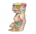
Animations - Stage 13
Movies - Embryo Carnegie stage 13 - These embryo animations rotate and show the relative position of internal systems at about the middle of embryonic development. Compare these with the later stage 22 embryos.The movies are based upon reconstruction of serial slice images.
|
|
|
|
Carnegie Stage 13 Movies: Gastrointestinal Tract | Cardiovascular | Central Nervous System | Carnegie Stage 13 | Stage 22 Movies | Movies
- The original animations were part of the UNSW Independent Learning Project (ILP) prepared by Aashish Kumar (2006).
Animations - Stage 22
Movies - Embryo Carnegie stage 22 - These embryo animations rotate and show the relative position of internal systems at the end of embryonic development. Compare these with the earlier stage 13 embryos. The movies are based upon reconstruction of serial slice images.
|
|
|
|
|
|
These 3d movies were part of the UNSW Medical degree Independent Learning Project (ILP) prepared by Aashish Kumar (2006).
Glossary Links
- Glossary: A | B | C | D | E | F | G | H | I | J | K | L | M | N | O | P | Q | R | S | T | U | V | W | X | Y | Z | Numbers | Symbols | Term Link
Cite this page: Hill, M.A. (2024, April 25) Embryology Embryo Serial Sections. Retrieved from https://embryology.med.unsw.edu.au/embryology/index.php/Embryo_Serial_Sections
- © Dr Mark Hill 2024, UNSW Embryology ISBN: 978 0 7334 2609 4 - UNSW CRICOS Provider Code No. 00098G