Developmental Signals - Wnt: Difference between revisions
No edit summary |
|||
| Line 18: | Line 18: | ||
==Function== | ==Function== | ||
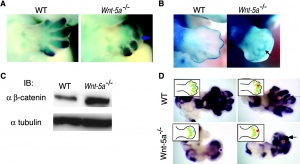
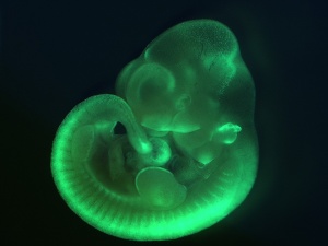
[[File:Mouse Wnt signaling 01.jpg|thumb|Mouse E10.5 Wnt signaling<ref name="PMID21176145"> | [[File:Mouse Wnt signaling 01.jpg|thumb|Mouse E10.5 Wnt signaling<ref name="PMID21176145">[http://www.ncbi.nlm.nih.gov/pubmed/21176145 PMID21176145]</ref>]] | ||
===Body Axis=== | ===Body Axis=== | ||
'''Wnt signaling and the polarity of the primary body axis'''<ref name="PMID20005801"><pubmed>20005801</pubmed></ref>"Data from diverse deuterostomes (frog, fish, mouse, and amphioxus) and from planarians (protostomes) suggest that Wnt signaling through beta-catenin controls posterior identity during body plan formation in most bilaterally symmetric animals." | '''Wnt signaling and the polarity of the primary body axis'''<ref name="PMID20005801"><pubmed>20005801</pubmed></ref>"Data from diverse deuterostomes (frog, fish, mouse, and amphioxus) and from planarians (protostomes) suggest that Wnt signaling through beta-catenin controls posterior identity during body plan formation in most bilaterally symmetric animals." | ||
Revision as of 14:15, 29 April 2011
Introduction

A secreted glycoprotein patterning switch with different roles in different tissues and signaling has generally been divided into the canonical and non-canonical pathways. The name was derived from two drosophila phenotypes wingless and int and the gene was first defined as a protooncogene, int1. Humans have 19 identified members, with the major subgroup of Wnts (WNT1, WNT3A, WNT8) signaling through activation of beta-catenin dependent transcription from at least 4 WNT genes encoding secreted glycoproteins. At least one wnt receptor called frizzled (FZD). Wnt7a is secreted protein and binds to extracellular matrix. The mechanism of Wnt distribution (free diffusion, restricted diffusion and active transport) and all its possible cell receptors are still being determined. In the gastrointestinal system, Wnt maintains the pool of undifferentiated intestinal progenitor cells and control maturation and correct positioning of the Paneth cell (a differentiated intestinal cell type).
The Wnt/beta-catenin signal pathway activation has also been identified in several cancers and appears to drive cell-cycle progression.
If you are interested in this family of proteins, look also at Roel Nusse -The Wnt Homepage.
| Factor Links: AMH | hCG | BMP | sonic hedgehog | bHLH | HOX | FGF | FOX | Hippo | LIM | Nanog | NGF | Nodal | Notch | PAX | retinoic acid | SIX | Slit2/Robo1 | SOX | TBX | TGF-beta | VEGF | WNT | Category:Molecular |
Some Recent Findings
|
Function

Body Axis
Wnt signaling and the polarity of the primary body axis[4]"Data from diverse deuterostomes (frog, fish, mouse, and amphioxus) and from planarians (protostomes) suggest that Wnt signaling through beta-catenin controls posterior identity during body plan formation in most bilaterally symmetric animals."
Neural

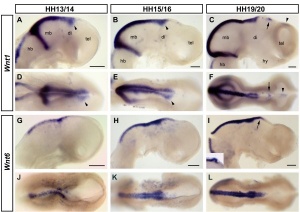
"Wnts are antagonised by Shh signalling. By demonstrating that Wnt4 expression in the thalamus is repressed by Shh from the ZLI we reveal an additional level of interaction between these two pathways and provide an example for the cross-regulation between patterning centres during forebrain regionalisation."[5]
Wnt receptor Frizzled-1 in presynaptic differentiation and function[6] "We examined the distribution of the Wnt receptor Frizzled-1 in cultured hippocampal neurons and determined that this receptor is located at synaptic contacts co-localizing with presynaptic proteins. Frizzled-1 was found in functional synapses detected with FM1-43 staining and in synaptic terminals from adult rat brain. Interestingly, overexpression of Frizzled-1 increased the number of clusters of Bassoon, a component of the active zone, while treatment with the extracellular cysteine-rich domain (CRD) of Frizzled-1 decreased Bassoon clustering, suggesting a role for this receptor in presynaptic differentiation. Consistent with this, treatment with the Frizzled-1 ligand Wnt-3a induced presynaptic protein clustering and increased functional presynaptic recycling sites, and these effects were prevented by co-treatment with the CRD of Frizzled-1."
(More? Neural System Development)
Muscle
Wnt signaling promotes AChR aggregation at the neuromuscular synapse[7]
- "Wnt proteins regulate the formation of central synapses by stimulating synaptic assembly, but their role at the vertebrate neuromuscular junction (NMJ) is unclear. Wnt3 is expressed by lateral motoneurons of the spinal cord during the period of motoneuron-muscle innervation. ...Wnt3 does not signal through the canonical Wnt pathway to induce cluster formation. Instead, Wnt3 induces the rapid formation of unstable AChR micro-clusters through activation of Rac1, which aggregate into large clusters only in the presence of agrin."
Vascular
Canonical Wnt signaling regulates organ-specific assembly and differentiation of CNS vasculature[8] "The central nervous system (CNS) vasculature consists of a tightly sealed endothelium that forms the blood-brain barrier, whereas blood vessels of other organs are more porous. Wnt7a and Wnt7b encode two Wnt ligands produced by the neuroepithelium of the developing CNS coincident with vascular invasion. Using genetic mouse models, we found that these ligands directly target the vascular endothelium and that the CNS uses the canonical Wnt signaling pathway to promote formation and CNS-specific differentiation of the organ's vasculature"
Signaling Pathway

At least 3 separate signaling pathways have been identified in relation to Wnt singnaling.
- Canonical pathway
- Non-canonical or planar cell polarity pathway
- Wnt-Calcium ion pathway
Canonical Pathway
- Wnt binds the surface receptor Frizzled (Fz) and LRP5/6 receptor complex
- Induces the stabilization of beta-catenin (through the DIX and PDZ domains of Dishevelled and other factors including Axin, glycogen synthase kinase 3 and casein kinase 1)
- Beta-catenin translocates into the nucleus
- Beta-catenin complexes with members of the LEF/TCF family of transcription factors.
- Transcriptional induction of target genes.
- Beta-catenin is then exported from the nucleus and degraded via the proteosomal machinery.
Non-Canonical Pathway
(Planar Cell Polarity Pathway)
- Wnt binds the surface receptor Frizzled (Fz) independent of the LRP5/6 receptor complex.
- Acts through Dishevelled (PDZ and DEP domains)
- Mediates cytoskeletal (microfilament) changes through activation of the small GTPases Rho and Rac.
Wnt-Calcium Ion Pathway
- Wnt binds the surface receptor Frizzled (Fz) and mediates activation of heterotrimeric G-proteins.
- Acts internally through Dsh, phospholipase C, calcium-calmodulin kinase 2 (CamK2) and protein kinase C (PKC).
- has various cellular functions including cell adhesion and motility.
Protein Data
- FUNCTION: PROBABLE DEVELOPMENTAL PROTEIN. SIGNALING BY WNT-7A ALLOWS SEXUALLY DIMORPHIC DEVELOPMENT OF THE MULLERIAN DUCTS (BY SIMILARITY).
- SUBCELLULAR LOCATION: POSSIBLY SECRETED AND ASSOCIATES WITH THE EXTRACELLULAR MATRIX.
- TISSUE SPECIFICITY: EXPRESSION IS RESTRICTED TO PLACENTA, KIDNEY, TESTIS, UTERUS, FETAL LUNG, AND FETAL AND ADULT BRAIN.
- SIMILARITY: BELONGS TO THE WNT FAMILY.
- FUNCTION: PROBABLE DEVELOPMENTAL PROTEIN. SIGNALING BY WNT-7A ALLOWS SEXUALLY DIMORPHIC DEVELOPMENT OF THE MULLERIAN DUCTS.
- SUBCELLULAR LOCATION: POSSIBLY SECRETED AND ASSOCIATES WITH THE EXTRACELLULAR MATRIX.
- DISEASE: MALE MICE LACKING WNT-7A FAIL TO UNDERGO REGRESSION OF THE MULLERIAN DUCT AS A RESULT OF THE ABSENCE OF THE RECEPTOR FOR MULLERIAN-INHIBITING SUBSTANCE. WNT7A-DEFICIENT FEMALES ARE INFERTILE BECAUSE OF ABNORMAL DEVELOPMENT OF THE OVIDUCT AND UTERUS, BOTH OF WHICH ARE MULLERIAN DUCT DERIVATIVES.
- SIMILARITY: BELONGS TO THE WNT FAMILY.
(data from Expasy)
OMIM
- WINGLESS-TYPE MMTV INTEGRATION SITE FAMILY, MEMBER 3A; WNT3A
- WINGLESS-TYPE MMTV INTEGRATION SITE FAMILY, MEMBER 7A; WNT7A
- CATENIN, BETA-1; CTNNB1
About OMIM "Online Mendelian Inheritance in Man OMIM is a comprehensive, authoritative, and timely compendium of human genes and genetic phenotypes. The full-text, referenced overviews in OMIM contain information on all known mendelian disorders and over 12,000 genes. OMIM focuses on the relationship between phenotype and genotype. It is updated daily, and the entries contain copious links to other genetics resources." OMIM
References
Reviews
<pubmed>20010950</pubmed> <pubmed>20005801</pubmed> <pubmed>19864126</pubmed> <pubmed>19736321</pubmed>
Search PubMed: Wnt
External Links
The nature of the internet is that some links may change over time. If the link no longer functions, search the internet using the link term.
- The Wnt gene Homepage by Roel Nusse
- Atlas of Genetics and Cytogenetics in Oncology and Haematology - The WNT Signaling Pathway and Its Role in Human Solid Tumors
- Mouse Wnt genes
- Comparative table of all vertebrate Wnt genes
- alignments of all mammalian Wnt genes
- Other Wnt pages
Glossary Links
- Glossary: A | B | C | D | E | F | G | H | I | J | K | L | M | N | O | P | Q | R | S | T | U | V | W | X | Y | Z | Numbers | Symbols | Term Link
Cite this page: Hill, M.A. (2024, April 18) Embryology Developmental Signals - Wnt. Retrieved from https://embryology.med.unsw.edu.au/embryology/index.php/Developmental_Signals_-_Wnt
- © Dr Mark Hill 2024, UNSW Embryology ISBN: 978 0 7334 2609 4 - UNSW CRICOS Provider Code No. 00098G