Developmental Signals - Homeobox: Difference between revisions
mNo edit summary |
mNo edit summary |
||
| Line 29: | Line 29: | ||
|-bgcolor="F5FAFF" | |-bgcolor="F5FAFF" | ||
| | | | ||
* '''Hox13 is essential for formation of a sensory organ at the terminal end of the sperm duct in Ciona'''{{#pmid:31682808|PMID31682808}} "Species-specific traits are thought to have been acquired by natural selection. Transcription factors play central roles in the evolution of species-specific traits. Hox genes encode a set of conserved transcription factors essential for establishing the anterior-posterior body axis of animals. Changes in the expression or function of Hox genes can lead to the diversification of animal-body plans. The tunicate ascidian Ciona intestinalis Type A has an orange-colored structure at the sperm duct terminus. This orange-pigmented organ (OPO) is the characteristic that can distinguish this ascidian from other closely related species. The OPO is formed by the accumulation of orange-pigmented cells (OPCs) that are present throughout the adult body. We show that Hox13 is essential for formation of the OPO. Hox13 is expressed in the epithelium of the sperm duct and neurons surrounding the terminal openings for sperm ejection, while OPCs themselves do not express this gene. OPCs are mobile cells that can move through the body vasculature by pseudopodia, suggesting that the OPO is formed by the accumulation of OPCs guided by Hox13-positive cells. Another ascidian species, Ciona savignyi, does not have an OPO. Like Hox13 of C. intestinalis, Hox13 of C. savignyi is expressed at the terminus of its sperm duct; however, its expression domain is limited to the circular area around the openings. The genetic changes responsible for the acquisition or loss of OPO are likely to occur in the expression pattern of Hox13." | |||
* '''Analysis of a limb-specific regulatory element in the promoter of the link protein gene'''{{#pmid:31470976|PMID31470976}} "Link protein is encoded by the Hapln1 gene and is a prototypical protein found in the cartilage matrix. It acts as an important component of the endochondral skeleton during early development. To study its transcriptional regulation, promoter fragments derived from the link protein gene were coupled to the β-galactosidase reporter and used to study in vivo transgene expression in mice. In day 15.5 mouse embryos, a link promoter fragment spanning -1020 to +40 nucleotides demonstrated highly specific β-galactosidase staining of skeletal structures, including the appendicular and axial cartilaginous tissues. Two shorter promoter fragments, spanning -690 to +40 and -315 to +40 nucleotides, demonstrated limb- and genitalia-specific expression resembling that of homeodomain-regulated tissues. Bioinformatic analysis revealed a highly conserved, Hox-like binding site (HLBS) at approximately -220 bp of the promoter, shared by both constructs, which contained the Hox-core consensus sequence TAATTA. Electromobility shift assays demonstrated binding of Hox-B4 recombinant protein to the HLBS, which was eliminated with nucleotide substitutions within the core-binding element. Co-transfection analysis of the HLBS demonstrated a 22-fold transcriptional activation by HoxA9 expression, which was ablated with a substitution within the core HLBS element. Together these findings establish promoter regions within the link protein gene that are important for in vivo expression and identify the potential role of homeodomain-containing proteins in controlling cartilage and limb gene expression." | |||
* '''A genome-wide assessment of the ancestral neural crest gene regulatory network'''{{#pmid:31619682|PMID31619682}} "The {{neural crest}} (NC) is an embryonic cell population that contributes to key vertebrate-specific features including the craniofacial skeleton and peripheral nervous system. Here we examine the transcriptional and epigenomic profiles of NC cells in the sea lamprey, in order to gain insight into the ancestral state of the NC gene regulatory network (GRN). Transcriptome analyses identify clusters of co-regulated genes during NC specification and migration that show high conservation across vertebrates but also identify transcription factors (TFs) and cell-adhesion molecules not previously implicated in NC migration. ATAC-seq analysis uncovers an ensemble of cis-regulatory elements, including enhancers of Tfap2B, SoxE1 and Hox-α2 validated in the embryo. Cross-species deployment of lamprey elements identifies the deep conservation of lamprey SoxE1 enhancer activity, mediating homologous expression in jawed vertebrates. Our data provide insight into the core GRN elements conserved to the base of the vertebrates and expose others that are unique to lampreys." | |||
* '''Hox genes in the pharyngeal region: how Hoxa3 controls early embryonic development of the pharyngeal organs'''{{#pmid:30604847|PMID30604847}} "The pharyngeal organs, namely the {{thyroid}}, {{thymus}}, {{parathyroid}}s, and ultimobranchial bodies, derive from the pharyngeal {{endoderm}} during embryonic development. The pharyngeal region is a segmented structure comprised of a series of reiterated structures: the pharyngeal arches on the exterior surface, the pharyngeal pouches on the interior, and a mesenchymal core. It is well known that Hox genes control spatial identity along the anterior-posterior axis of the developing vertebrate embryo, and nowhere is this is more evident than in the pharyngeal region. Each of the distinct segmented regions has a unique pattern of Hox expression, which conveys crucial positional information to the cells and tissues within it. In the context of pharyngeal organ development, molecular data suggest that HOXA3 is responsible for specifying organ identity within the third pharyngeal pouch, and in its absence, thymus and parathyroid organogenesis fails to proceed normally" | * '''Hox genes in the pharyngeal region: how Hoxa3 controls early embryonic development of the pharyngeal organs'''{{#pmid:30604847|PMID30604847}} "The pharyngeal organs, namely the {{thyroid}}, {{thymus}}, {{parathyroid}}s, and ultimobranchial bodies, derive from the pharyngeal {{endoderm}} during embryonic development. The pharyngeal region is a segmented structure comprised of a series of reiterated structures: the pharyngeal arches on the exterior surface, the pharyngeal pouches on the interior, and a mesenchymal core. It is well known that Hox genes control spatial identity along the anterior-posterior axis of the developing vertebrate embryo, and nowhere is this is more evident than in the pharyngeal region. Each of the distinct segmented regions has a unique pattern of Hox expression, which conveys crucial positional information to the cells and tissues within it. In the context of pharyngeal organ development, molecular data suggest that HOXA3 is responsible for specifying organ identity within the third pharyngeal pouch, and in its absence, thymus and parathyroid organogenesis fails to proceed normally" | ||
Revision as of 07:42, 7 November 2019
| Embryology - 25 Apr 2024 |
|---|
| Google Translate - select your language from the list shown below (this will open a new external page) |
|
العربية | català | 中文 | 中國傳統的 | français | Deutsche | עִברִית | हिंदी | bahasa Indonesia | italiano | 日本語 | 한국어 | မြန်မာ | Pilipino | Polskie | português | ਪੰਜਾਬੀ ਦੇ | Română | русский | Español | Swahili | Svensk | ไทย | Türkçe | اردو | ייִדיש | Tiếng Việt These external translations are automated and may not be accurate. (More? About Translations) |
Introduction

The family of homeobox (Hox) proteins has been a focus of research for over 30 years. In humans, the homeobox gene family contains about 235 functional genes and 65 pseudogenes. This family of genes were also the basis of the embryo patterning studies that led to the Nobel Prize in Medicine 1995. We now know that in addition to whole embryo axes patterning, this family of genes has many roles in establishing pattern throughout the embryo in different tissues and organs.
There has recently been a revival of an earlier theory[2] that Hox expression during vertebrate pattern formation is linked to the process of segmentation of paraxial mesoderm during somitogenesis.[3]
This signalling pathway has also been implicated in many developmental abnormalities and diseases.

|

|
| Fly wild-type head{{pmid:108157|PMID108157}} | Fly antennapedia head{{pmid:108157|PMID108157}} |
| Factor Links: AMH | hCG | BMP | sonic hedgehog | bHLH | HOX | FGF | FOX | Hippo | LIM | Nanog | NGF | Nodal | Notch | PAX | retinoic acid | SIX | Slit2/Robo1 | SOX | TBX | TGF-beta | VEGF | WNT | Category:Molecular |
Some Recent Findings


|
| More recent papers |
|---|
 This table allows an automated computer search of the external PubMed database using the listed "Search term" text link.
More? References | Discussion Page | Journal Searches | 2019 References | 2020 References Search term: Hox | antennapedia |
| Older papers |
|---|
| These papers originally appeared in the Some Recent Findings table, but as that list grew in length have now been shuffled down to this collapsible table.
See also the Discussion Page for other references listed by year and References on this current page.
|
Classification
|
|
| Proposed Hox protein classification[1] |
Human Hox

Chromosomal Distribution of Human Homeobox Genes[16]
Functions
Developmental patterning signal.
Neural
Segmentation

|
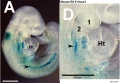
Hindbrain neural crest migration and Hox expression pattern[17]
Legend
Adapted by permission from Macmillan Publishers Ltd: Nature Reviews Neuroscience[17], copyright (2007) |
Phrenic Motor Neurons
Hox5 (Hoxa5 and Hoxc5) required for phrenic motor column (PMC) development that form the respiratory motor neurons driving the diaphragm for respiration.[18]
Axial Skeleton

Vertebral element ossification between species.[19] |

Chicken model of Hox in paraxial mesoderm precursors in the epiblast/tail-bud during axis elongation[20] |
- Links: Somitogenesis | Axial Skeleton Development
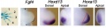
Limb

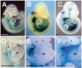
Mouse Limb Patterning Fgf and Hox Expression[21]
Fgf and Hox expression in E10.5 to 10.75 wild-type embryonic forelimb autopod, compared to future E14.5 digit arrangement.

Expression of Hoxa4, Hoxa9, Hoxa10, Hoxa11, Hoxa11 antisense (Hoxa11as), and Hoxa13 in E12.5 limb buds.[22]
- Links: Limb Development
Other
Signaling Pathway
Otx
Otx is a txanscription factor essential for the normal development of the brain, cerebellum, pineal gland, and eye.
OTX is a homeobox family gene related to a gene expressed in the developing Drosophila head termed 'orthodenticle.' OTX transcription factors bind with high affinity to TAATCC/T elements on DNA.
- Links: Pineal | Vision | PubMed - Otx
Additional Images
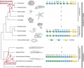
Hox deuterostomes phylogenetic tree PMID 23819519
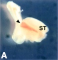
Ascidian hox expression
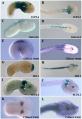
Hoxa3 Mouse E8.5-E10.5
Hoxa3 Mouse E8.5
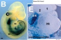
Hoxa3 Mouse E9.5
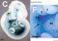
Hoxa3 Mouse E10.5
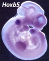
Hoxb5 Mouse
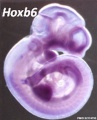
Hoxb6 Mouse
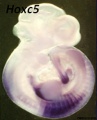
Hoxc5 Mouse
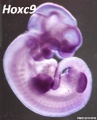
Hoxc9 Mouse
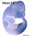
Hoxc10 Mouse
Mouse forelimb bud Fgf4-Hoxd13-Hoxa13
Mouse spleen Hox11 expression


References
- ↑ 1.0 1.1 1.2 Hueber SD, Weiller GF, Djordjevic MA & Frickey T. (2010). Improving Hox protein classification across the major model organisms. PLoS ONE , 5, e10820. PMID: 20520839 DOI.
- ↑ Meinhardt H. Models For Biological Pattern Formation. Academic Press; 1982.
- ↑ Gu S, Gu W, Shou J, Xiong J, Liu X, Sun B, Yang D & Xie R. (2017). The Molecular Feature of HOX Gene Family in the Intramedullary Spinal Tumors. Spine , 42, 291-297. PMID: 25785959 DOI.
- ↑ 4.0 4.1 Coulombe Y, Lemieux M, Moreau J, Aubin J, Joksimovic M, Bérubé-Simard FA, Tabariès S, Boucherat O, Guillou F, Larochelle C, Tuggle CK & Jeannotte L. (2010). Multiple promoters and alternative splicing: Hoxa5 transcriptional complexity in the mouse embryo. PLoS ONE , 5, e10600. PMID: 20485555 DOI.
- ↑ Tajima Y, Hozumi A, Yoshida K, Treen N, Sakuma T, Yamamoto T & Sasakura Y. (2019). Hox13 is essential for formation of a sensory organ at the terminal end of the sperm duct in Ciona. Dev. Biol. , , . PMID: 31682808 DOI.
- ↑ Rhodes CS, Matsunobu T & Yamada Y. (2019). Analysis of a limb-specific regulatory element in the promoter of the link protein gene. Biochem. Biophys. Res. Commun. , 518, 672-677. PMID: 31470976 DOI.
- ↑ Hockman D, Chong-Morrison V, Green SA, Gavriouchkina D, Candido-Ferreira I, Ling ITC, Williams RM, Amemiya CT, Smith JJ, Bronner ME & Sauka-Spengler T. (2019). A genome-wide assessment of the ancestral neural crest gene regulatory network. Nat Commun , 10, 4689. PMID: 31619682 DOI.
- ↑ Gordon J. (2018). Hox genes in the pharyngeal region: how Hoxa3 controls early embryonic development of the pharyngeal organs. Int. J. Dev. Biol. , 62, 775-783. PMID: 30604847 DOI.
- ↑ Acemel RD, Tena JJ, Irastorza-Azcarate I, Marlétaz F, Gómez-Marín C, de la Calle-Mustienes E, Bertrand S, Diaz SG, Aldea D, Aury JM, Mangenot S, Holland PW, Devos DP, Maeso I, Escrivá H & Gómez-Skarmeta JL. (2016). A single three-dimensional chromatin compartment in amphioxus indicates a stepwise evolution of vertebrate Hox bimodal regulation. Nat. Genet. , 48, 336-41. PMID: 26829752 DOI.
- ↑ Hench J, Henriksson J, Abou-Zied AM, Lüppert M, Dethlefsen J, Mukherjee K, Tong YG, Tang L, Gangishetti U, Baillie DL & Bürglin TR. (2015). The Homeobox Genes of Caenorhabditis elegans and Insights into Their Spatio-Temporal Expression Dynamics during Embryogenesis. PLoS ONE , 10, e0126947. PMID: 26024448 DOI.
- ↑ Parker HJ, Bronner ME & Krumlauf R. (2014). A Hox regulatory network of hindbrain segmentation is conserved to the base of vertebrates. Nature , 514, 490-3. PMID: 25219855 DOI.
- ↑ Zigman M, Laumann-Lipp N, Titus T, Postlethwait J & Moens CB. (2014). Hoxb1b controls oriented cell division, cell shape and microtubule dynamics in neural tube morphogenesis. Development , 141, 639-49. PMID: 24449840 DOI.
- ↑ Natale A, Sims C, Chiusano ML, Amoroso A, D'Aniello E, Fucci L, Krumlauf R, Branno M & Locascio A. (2011). Evolution of anterior Hox regulatory elements among chordates. BMC Evol. Biol. , 11, 330. PMID: 22085760 DOI.
- ↑ Yallowitz AR, Hrycaj SM, Short KM, Smyth IM & Wellik DM. (2011). Hox10 genes function in kidney development in the differentiation and integration of the cortical stroma. PLoS ONE , 6, e23410. PMID: 21858105 DOI.
- ↑ Zhang Y, Huang Q, Cheng JC, Nishi Y, Yanase T, Huang HF & Leung PC. (2010). Homeobox A7 increases cell proliferation by up-regulation of epidermal growth factor receptor expression in human granulosa cells. Reprod. Biol. Endocrinol. , 8, 61. PMID: 20540809 DOI.
- ↑ Holland PW, Booth HA & Bruford EA. (2007). Classification and nomenclature of all human homeobox genes. BMC Biol. , 5, 47. PMID: 17963489 DOI.
- ↑ 17.0 17.1 Guthrie S. (2007). Patterning and axon guidance of cranial motor neurons. Nat. Rev. Neurosci. , 8, 859-71. PMID: 17948031 DOI.
- ↑ Philippidou P, Walsh CM, Aubin J, Jeannotte L & Dasen JS. (2012). Sustained Hox5 gene activity is required for respiratory motor neuron development. Nat. Neurosci. , 15, 1636-44. PMID: 23103965 DOI.
- ↑ Hautier L, Weisbecker V, Sánchez-Villagra MR, Goswami A & Asher RJ. (2010). Skeletal development in sloths and the evolution of mammalian vertebral patterning. Proc. Natl. Acad. Sci. U.S.A. , 107, 18903-8. PMID: 20956304 DOI.
- ↑ Denans N, Iimura T & Pourquié O. (2015). Hox genes control vertebrate body elongation by collinear Wnt repression. Elife , 4, . PMID: 25719209 DOI.
- ↑ Galli A, Robay D, Osterwalder M, Bao X, Bénazet JD, Tariq M, Paro R, Mackem S & Zeller R. (2010). Distinct roles of Hand2 in initiating polarity and posterior Shh expression during the onset of mouse limb bud development. PLoS Genet. , 6, e1000901. PMID: 20386744 DOI.
- ↑ Woltering JM, Noordermeer D, Leleu M & Duboule D. (2014). Conservation and divergence of regulatory strategies at Hox Loci and the origin of tetrapod digits. PLoS Biol. , 12, e1001773. PMID: 24465181 DOI.
Reviews
Mallo M, Wellik DM & Deschamps J. (2010). Hox genes and regional patterning of the vertebrate body plan. Dev. Biol. , 344, 7-15. PMID: 20435029 DOI.
Narita Y & Rijli FM. (2009). Hox genes in neural patterning and circuit formation in the mouse hindbrain. Curr. Top. Dev. Biol. , 88, 139-67. PMID: 19651304 DOI.
Deschamps J & van Nes J. (2005). Developmental regulation of the Hox genes during axial morphogenesis in the mouse. Development , 132, 2931-42. PMID: 15944185 DOI.
Schilling TF & Knight RD. (2001). Origins of anteroposterior patterning and Hox gene regulation during chordate evolution. Philos. Trans. R. Soc. Lond., B, Biol. Sci. , 356, 1599-613. PMID: 11604126 DOI.
Articles
Search Pubmed
Search Bookshelf hox
Search Pubmed Now: Hox Homeobox
External Links
External Links Notice - The dynamic nature of the internet may mean that some of these listed links may no longer function. If the link no longer works search the web with the link text or name. Links to any external commercial sites are provided for information purposes only and should never be considered an endorsement. UNSW Embryology is provided as an educational resource with no clinical information or commercial affiliation.
- Nobel Prize - Medicine 1995
- OMIM - SONIC HEDGEHOG
- Genetics Home Reference
Glossary Links
- Glossary: A | B | C | D | E | F | G | H | I | J | K | L | M | N | O | P | Q | R | S | T | U | V | W | X | Y | Z | Numbers | Symbols | Term Link
Cite this page: Hill, M.A. (2024, April 25) Embryology Developmental Signals - Homeobox. Retrieved from https://embryology.med.unsw.edu.au/embryology/index.php/Developmental_Signals_-_Homeobox
- © Dr Mark Hill 2024, UNSW Embryology ISBN: 978 0 7334 2609 4 - UNSW CRICOS Provider Code No. 00098G