Introduction
Draft Page - notice removed when complete.
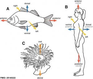
Anatomical axes comparison
How do you establish the anatomical axes of the embryo? Another well studied model of axis patterning is the establishment of limb axes, in particular this system historically was studied by grafting and rotating parts of the early developing limb.
Note there is some confusion arising in the terminology when comparing animal developmental axes and those of human anatomical axes.
Left-right axis (L-R) transverse plane
- vertebrates - left side Nodal expression
- sea urchins - right side Nodal expression, inhibit the right mesodermal coelomic pouch (CP) from forming the adult rudiment
Signaling Factors
- Nodal - induces its own expression and that of Lefty.
- Lefty - a Nodal feedback inhibitor with a longer diffusion range.
|
|
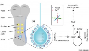
| Primitive node cilia (human stage 8)
|
Embryo left-right asymmetry pathway[1]
|
Axes Formation: Dorsal-Ventral Axis | Cranio-Caudal Axis | Template:Left-Right Axis
Some Recent Findings
- zebrafish znfl1s regulate left-right asymmetry patterning through controlling the expression of fgfr1a[2] "Proper left-right (LR) axis establishment is critical for organogenesis in vertebrates. Previously, we reported that zinc finger transcription factors zinc finger transcription factor 1 (znfl1s) are expressed in the tailbud and axial mesoderm in zebrafish. However, a role of znfl1s in LR axis development has not been demonstrated. Here, we discovered that the knockdown of znfl1s using morpholino (MO) in whole embryos or dorsal forerunner cells (DFCs) interrupted LR asymmetry and normal development of the heart, liver, and pancreas. Whole-embryo knockdown of znfl1s by MO or clustered regularly interspaced short palindromic repeat (CRISPR) interference (CRISPRi) resulted in the absent expression of nodal gene spaw and Nodal signaling-related genes lft1, lft2, and pitx2c in the left lateral plate mesoderm (LPM), and Spaw, Lft1, Lft2, and Pitx2c play important roles in LR axis development in zebrafish. However, specific knockdown of znfl1s in DFCs resulted in random expression of spaw, lft1, lft2, and pitx2c. Knockdown of znfl1s led to abnormal cilia formation by the downregulation of fgfr1a and foxj1a expression. The expression of spaw, lft1, lft2, and pitx2c was partially rescued by the overexpression of fgfr1a mRNA in znfl1s morphants. Taken together, our results suggest that znfl1s regulate laterality development in zebrafish embryos through controlling the expression of fgfr1a."
- Paraxial Nodal Expression Reveals a Novel Conserved Structure of the Left-Right Organizer in Four Mammalian Species[3] "Nodal activity in the left lateral plate mesoderm is a conserved sign of irreversible left-right asymmetry at early somite stages of the vertebrate embryo. An earlier, paraxial nodal domain accompanies the emergence and initial extension of the notochord and is either left-sided, as in the chick and pig, or symmetrical, as in the mouse and rabbit; intriguingly, this interspecific dichotomy is mirrored by divergent morphological features of the posterior notochord (also known as the left-right organizer), which is ventrally exposed to the yolk sac cavity and carries motile cilia in the latter 2 species only. By introducing the cattle embryo as a new model organism for early left-right patterning, we present data to establish 2 groups of mammals characterized by both the morphology of the left-right organizer and the dynamics of paraxial nodal expression: presence and absence of a ventrally open surface of the early (plate-like) posterior notochord correlates with a symmetrical (in mice and rabbits) versus an asymmetrical (in pigs and cattle) paraxial nodal expression domain next to the notochordal plate. High-resolution histological analysis reveals that the latter domain defines in all 4 mammals a novel 'parachordal' axial mesoderm compartment, the topography of which changes according to the specific regression of the similarly novel subchordal mesoderm during the initial phases of notochord development. In conclusion, the mammalian axial mesoderm compartment (1) shares critical conserved features despite the marked differences in early notochord morphology and early left-right patterning and (2) provides a dynamic topographical framework for nodal activity as part of the mammalian left-right organiser."
|
| More recent papers
|
|
This table allows an automated computer search of the external PubMed database using the listed "Search term" text link.
- This search now requires a manual link as the original PubMed extension has been disabled.
- The displayed list of references do not reflect any editorial selection of material based on content or relevance.
- References also appear on this list based upon the date of the actual page viewing.
References listed on the rest of the content page and the associated discussion page (listed under the publication year sub-headings) do include some editorial selection based upon both relevance and availability.
More? References | Discussion Page | Journal Searches | 2019 References | 2020 References
Search term: Left-Right Axis
|
Nodal
Nodal is expressed during mouse gastrulation and is a member of the TGF-beta gene family.
- Links: OMIM601265
Lefty1
Lefty1 and Lefty2 are expressed in developing mouse embryos on the left side. These are members of the TGF-beta gene family.
LEFTY A and LEFTY B are the human homologues of murine genes implicated in left-right axis development.
- Links: OMIM603037
References
- ↑ Norris DP. (2012). Cilia, calcium and the basis of left-right asymmetry. BMC Biol. , 10, 102. PMID: 23256866 DOI.
- ↑ Li J, Gao F, Zhao Y, He L, Huang Y, Yang X, Zhou Y, Yu L, Zhao Q & Dong X. (2019). Zebrafish znfl1s regulate left-right asymmetry patterning through controlling the expression of fgfr1a. J. Cell. Physiol. , 234, 1987-1995. PMID: 30317609 DOI.
- ↑ Li J, Gao F, Zhao Y, He L, Huang Y, Yang X, Zhou Y, Yu L, Zhao Q & Dong X. (2019). Zebrafish znfl1s regulate left-right asymmetry patterning through controlling the expression of fgfr1a. J. Cell. Physiol. , 234, 1987-1995. PMID: 30317609 DOI.
Reviews
Articles
Glossary Links
- Glossary: A | B | C | D | E | F | G | H | I | J | K | L | M | N | O | P | Q | R | S | T | U | V | W | X | Y | Z | Numbers | Symbols | Term Link
Cite this page: Hill, M.A. (2024, April 16) Embryology Developmental Mechanism - Left-Right Axis. Retrieved from https://embryology.med.unsw.edu.au/embryology/index.php/Developmental_Mechanism_-_Left-Right_Axis
- What Links Here?
- © Dr Mark Hill 2024, UNSW Embryology ISBN: 978 0 7334 2609 4 - UNSW CRICOS Provider Code No. 00098G


