Detailed Cardiac - Sinus Node

From Embryology
Embryology - 16 Apr 2024    Facebook link Pinterest link Twitter link  Expand to Translate  
Google Translate - select your language from the list shown below (this will open a new external page)

العربية | català | 中文 | 中國傳統的 | français | Deutsche | עִברִית | हिंदी | bahasa Indonesia | italiano | 日本語 | 한국어 | မြန်မာ | Pilipino | Polskie | português | ਪੰਜਾਬੀ ਦੇ | Română | русский | Español | Swahili | Svensk | ไทย | Türkçe | اردو | ייִדיש | Tiếng Việt    These external translations are automated and may not be accurate. (More? About Translations)

Online Editor 
Mark Hill.jpg
This content was contributed by Prof Robert H. Anderson.


Anderson RH. Teratogenecity in the setting of cardiac development and maldevelopment. (2016)

Please note that the content is still at the early draft stage and will be the basis of updating the online cardiac development information with a more detailed description as shown by the page links below (shown in red if not yet existing).

Detailed Cardiac: Systemic Venous Sinus | Pulmonary Vein | Superior Interatrial Fold | Atrioventricular Cushions | Atrioventricular Canal | Interventricular Communication | Subpulmonary Infundibulum | Arterial Roots | Intrapericardial Arterial Trunks | Extrapericardial Arterial Channels | Sinus Node | Atrioventricular Conduction Axis


Cardiovascular Links: cardiovascular | Heart Tutorial | Lecture - Early Vascular | Lecture - Heart | Movies | 2016 Cardiac Review | heart | coronary circulation | heart valve | heart rate | Circulation | blood | blood vessel | blood vessel histology | heart histology | Lymphatic | ductus venosus | spleen | Stage 22 | cardiovascular abnormalities | OMIM | 2012 ECHO Meeting | Category:Cardiovascular
Historic Embryology - Cardiovascular 
1902 Vena cava inferior | 1905 Brain Blood Vessels | 1909 Cervical Veins | 1909 Dorsal aorta and umbilical veins | 1912 Heart | 1912 Human Heart | 1914 Earliest Blood-Vessels | 1915 Congenital Cardiac Disease | 1915 Dura Venous Sinuses | 1916 Blood cell origin | 1916 Pars Membranacea Septi | 1919 Lower Limb Arteries | 1921 Human Brain Vascular | 1921 Spleen | 1922 Aortic-Arch System | 1922 Pig Forelimb Arteries | 1922 Chicken Pulmonary | 1923 Head Subcutaneous Plexus | 1923 Ductus Venosus | 1925 Venous Development | 1927 Stage 11 Heart | 1928 Heart Blood Flow | 1935 Aorta | 1935 Venous valves | 1938 Pars Membranacea Septi | 1938 Foramen Ovale | 1939 Atrio-Ventricular Valves | 1940 Vena cava inferior | 1940 Early Hematopoiesis | 1941 Blood Formation | 1942 Truncus and Conus Partitioning | Ziegler Heart Models | 1951 Heart Movie | 1954 Week 9 Heart | 1957 Cranial venous system | 1959 Brain Arterial Anastomoses | Historic Embryology Papers | 2012 ECHO Meeting | 2016 Cardiac Review | Historic Disclaimer


Search: PubMed - Anderson RH

Detailed Cardiac: Systemic Venous Sinus | Pulmonary Vein | Superior Interatrial Fold | Atrioventricular Cushions | Atrioventricular Canal | Interventricular Communication | Subpulmonary Infundibulum | Arterial Roots | Intrapericardial Arterial Trunks | Extrapericardial Arterial Channels | Sinus Node | Atrioventricular Conduction Axis

Appearance and Remodelling of the Sinus Node

Introduction

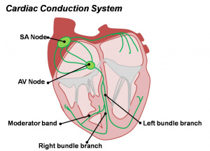
Cardiac Conduction System

The sinoatrial node (SAN) is the primary pacemaker of the heart that initially develops from part of pro-pacemaking embryonic venous pole that expresses both Hcn4 and the transcriptional factor Shox2.[1] The adult node is a mixture of about 10,000 cells distinct myocytes located at the entry of the right superior caval vein (SVC) into the right atrium.


Cardiovascular Links: cardiovascular | Heart Tutorial | Lecture - Early Vascular | Lecture - Heart | Movies | 2016 Cardiac Review | heart | coronary circulation | heart valve | heart rate | Circulation | blood | blood vessel | blood vessel histology | heart histology | Lymphatic | ductus venosus | spleen | Stage 22 | cardiovascular abnormalities | OMIM | 2012 ECHO Meeting | Category:Cardiovascular
Historic Embryology - Cardiovascular 
1902 Vena cava inferior | 1905 Brain Blood Vessels | 1909 Cervical Veins | 1909 Dorsal aorta and umbilical veins | 1912 Heart | 1912 Human Heart | 1914 Earliest Blood-Vessels | 1915 Congenital Cardiac Disease | 1915 Dura Venous Sinuses | 1916 Blood cell origin | 1916 Pars Membranacea Septi | 1919 Lower Limb Arteries | 1921 Human Brain Vascular | 1921 Spleen | 1922 Aortic-Arch System | 1922 Pig Forelimb Arteries | 1922 Chicken Pulmonary | 1923 Head Subcutaneous Plexus | 1923 Ductus Venosus | 1925 Venous Development | 1927 Stage 11 Heart | 1928 Heart Blood Flow | 1935 Aorta | 1935 Venous valves | 1938 Pars Membranacea Septi | 1938 Foramen Ovale | 1939 Atrio-Ventricular Valves | 1940 Vena cava inferior | 1940 Early Hematopoiesis | 1941 Blood Formation | 1942 Truncus and Conus Partitioning | Ziegler Heart Models | 1951 Heart Movie | 1954 Week 9 Heart | 1957 Cranial venous system | 1959 Brain Arterial Anastomoses | Historic Embryology Papers | 2012 ECHO Meeting | 2016 Cardiac Review | Historic Disclaimer

Some Recent Findings

  • Computational analysis of the human sinus node action potential: model development and effects of mutations[2] "The sinoatrial node (SAN) is the normal pacemaker of the mammalian heart. Over several decades, a large amount of data on the ionic mechanisms underlying the spontaneous electrical activity of SAN pacemaker cells has been obtained, mostly in experiments on single cells isolated from rabbit SAN. This wealth of data has allowed the development of mathematical models of the electrical activity of rabbit SAN pacemaker cells. Our aim was to construct a more comprehensive model of the electrical activity of a human SAN pacemaker cell, using recently obtained electrophysiological data from human SAN pacemaker cells. We based our model on the recent Severi-DiFrancesco model of a rabbit SAN pacemaker cell. The action potential and calcium transient of the resulting model are close to the experimentally recorded values: the model has a much smaller 'funny current' (If ) than do rabbit cells, but its modulatory role is highly similar. Changes in pacing rate upon the implementation of mutations associated with sinus node dysfunction agree with the clinical observations. This agreement holds for both loss-of-function and gain-of-function mutations in the HCN4, SCN5A, and KCNQ1 genes, underlying ion channelopathies in If , fast sodium current, and slow delayed rectifier potassium current, respectively. We conclude that our human SAN cell model can be a useful tool in the design of experiments and the development of drugs that aim to modulate heart rate.
  • Development of the cardiac pacemaker[3] "The sinoatrial node (SAN) is the dominant pacemaker of the heart. Abnormalities in SAN formation and function can cause sinus arrhythmia, including sick sinus syndrome and sudden death. A better understanding of genes and signaling pathways that regulate SAN development and function is essential to develop more effective treatment to sinus arrhythmia, including biological pacemakers. In this review, we briefly summarize the key processes of SAN morphogenesis during development, and focus on the transcriptional network that drives SAN development."
More recent papers  
Mark Hill.jpg
PubMed logo.gif

This table allows an automated computer search of the external PubMed database using the listed "Search term" text link.

  • This search now requires a manual link as the original PubMed extension has been disabled.
  • The displayed list of references do not reflect any editorial selection of material based on content or relevance.
  • References also appear on this list based upon the date of the actual page viewing.


References listed on the rest of the content page and the associated discussion page (listed under the publication year sub-headings) do include some editorial selection based upon both relevance and availability.

More? References | Discussion Page | Journal Searches | 2019 References | 2020 References

Search term: Sinus Node Development

<pubmed limit=5>Sinus Node Development</pubmed>

Molecular

Hcn4

Hyperpolarization-Activated Cyclic Nucleotide-Gated Potassium Channel 4 (HCN4) gene located at 15q24.1, the 1,203 amino acid protein contains 6 putative transmembrane segments, a pore region, and a cyclic nucleotide-binding domain.

Links: [OMIM HCN4

Shox2

Short Stature Homeobox 2 (SHOX2) gene located at 3q25.32, the protein acts as a homeobox transcription factor.

  • mouse og12 gene - developmental sinus venosus (aorta), female genitalia, diencephalon, mes- and myelencephalon, nasal capsula, palate, eyelid, and limbs
Links: OMIM SHOX2

ISL1

ISL Lim Homeobox 1 (ISL1) gene located at 5q11.1 is a member of the LIM/homeodomain family of transcription factors that binds to the enhancer region of the insulin gene. Functions in survival, proliferation, and function of pacemaker cells throughout development.[4]


Links: OMIM ISL1

References

  1. <pubmed>26682210</pubmed>
  2. <pubmed>28185290</pubmed>
  3. <pubmed>27770149</pubmed>
  4. <pubmed>26193633</pubmed>

Reviews

<pubmed>26023305</pubmed> <pubmed>24672485</pubmed> <pubmed></pubmed> <pubmed></pubmed>

Articles

<pubmed>27291063</pubmed> <pubmed>26682210</pubmed> <pubmed>26611337</pubmed> <pubmed>26835096</pubmed>


Cite this page: Hill, M.A. (2024, April 16) Embryology Detailed Cardiac - Sinus Node. Retrieved from https://embryology.med.unsw.edu.au/embryology/index.php/Detailed_Cardiac_-_Sinus_Node

What Links Here?
© Dr Mark Hill 2024, UNSW Embryology ISBN: 978 0 7334 2609 4 - UNSW CRICOS Provider Code No. 00098G